- FSH-receptor
-
The follicle-stimulating hormone receptor or FSH-receptor (FSHR) is a transmembrane receptor that interacts with the follicle-stimulating hormone (FSH) and represents a G protein-coupled receptor (GPCR). Its activation is necessary for the hormonal functioning of FSH. FSHRs are found in the ovary, testis, and uterus.
Contents
FSHR gene
The gene for the FSHR is found on chromosome 2 p21 in humans. The gene sequence of the FSHR consists of about 2,080 nucleotides.[1]
Receptor structure
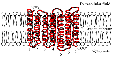
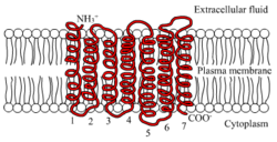 The seven transmembrane α-helix structure of a G protein-coupled receptor such as FSHR
The FSHR consists of 695 amino acids and has a molecular mass of about 76 kDa.[1] Like other GPCRs, the FSH-receptor possesses seven membrane-spanning domains or transmembrane helices.
- The extracellular domain of the receptor is glycosylated.
- The transmembrane domain contains two highly conserved cysteine residues that build disulfide bonds to stabilize the receptor structure. A highly conserved Asp-Arg-Tyr triplet motif is present and may be of importance to transmit the signal.
- The C-terminal domain is intracellular and brief, rich in serine and threonine residues for possible phosphorylation.
Ligand binding and signal transduction
Upon binding FSH externally to the membrane, a transduction of the signal that activates the G protein that is bound to the receptor internally takes place. With FSH attached, the receptor shifts conformation and, thus, mechanically activates the G protein, which detaches from the receptor and activates the cAMP system.
It is believed that a receptor molecule exists in a conformational equilibrium between active and inactive states. The binding of FSH to the receptor shifts the equilibrium between active and inactive receptors. FSH and FSH-agonists shift the equilibrium in favor of active states; FSH antagonists shift the equilibrium in favor of inactive states. For a cell to respond to FSH, only a small percentage (~1%) of receptor sites need to be activated.
Phosphorylation by cAMP-dependent protein kinases
Cyclic AMP-dependent protein kinases (protein kinase A) are activated by the signal chain coming from the G protein (that was activated by the FSH-receptor) via adenylate cyclase and cyclic AMP (cAMP).
These protein kinases are present as tetramers with two regulatory units and two catalytic units. Upon binding of cAMP to the regulatory units, the catalytic units are released and initiate the phosphorylation of proteins, leading to the physiologic action. The cyclic AMP-regulatory dimers are degraded by phosphodiesterase and release 5’AMP. DNA in the cell nucleus binds to phosphorylated proteins through the cyclic AMP response element (CRE), which results in the activation of genes.[1]The signal is amplified by the involvement of cAMP and the resulting phosphorylation. The process is modified by prostaglandins. Other cellular regulators are participate are the intracellular calcium concentration modified by phospholipase, nitric acid, and other growth factors.
The FSH receptor can also activate the extracellular signal-regulated kinases (ERK).[2] In a feedback mechanism, these activated kinases phosphorylate the receptor. The longer the receptor remains active, the more kinases are activated, the more receptors are phosphorylated.
Action
In the ovary, the FSH receptor is necessary for follicular development and expressed on the granulosa cells.[1]
In the male, the FSH receptor has been identified on the Sertoli cells that are critical for spermatogenesis.[3]
The FSHR is expressed during the luteal phase in the secretory endometrium of the uterus.[4]
FSH receptor is selectively expressed on the surface of the blood vessels of a wide range of carcinogenic tumors.[5]
Receptor regulation
Upregulation
Upregulation refers to the increase in the number of receptor site on the membrane. Estrogen upregulates FSH receptor sites. In turn, FSH stimulates granulosa cells to produce estrogens. This synergistic activity of estrogen and FSH allows for follicle growth and development in the ovary.
Desensitization
The FSHR become desensitized when exposed to FSH for some time. A key reaction of this downregulation is the phosphorylation of the intracellular (or cytoplasmic) receptor domain by protein kinases. This process uncouples Gs protein from the FSHR. Another way to desensitize is to uncouple the regulatory and catalytic units of the cAMP system.
Downregulation
Downregulation refers to the decrease in the number of receptor sites. This can be accomplished by metabolizing bound FSHR sites. The bound FSH-receptor complex is brought by lateral migration to a "coated pit," where such units are concentrated and then stabilized by a framework of clathrins. A pinched-off coated pit is internalized and degraded by lysosomes. Proteins may be metabolized or the receptor can be recycled. Use of long-acting agonists will downregulate the receptor population.
Modulators
Antibodies to FSHR can interfere with FSHR activity.
FSHR abnormalities
Some patients with ovarian hyperstimulation syndrome may have mutations in the gene for FSHR, making them more sensitive to gonadotropin stimulation.[6]
Women with 46 XX gonadal dysgenesis experience primary amenorrhea with hypergonadotropic hypogonadism. There are forms of 46 xx gonadal dysgenesis wherein abnormalities in the FSH-receptor have been reported and are thought to be the cause of the hypogonadism.[7]
Polymorphism may affect FSH receptor populations and lead to poorer responses in infertile women receiving FSH medication for IVF.[8]
History
Alfred G. Gilman and Martin Rodbell received the 1994 Nobel Prize in Medicine and Physiology for the discovery of the G Protein System.
See also
References
- ^ a b c d Simoni M, Gromoll J, Nieschlag E (December 1997). "The follicle-stimulating hormone receptor: biochemistry, molecular biology, physiology, and pathophysiology". Endocr. Rev. 18 (6): 739–73. doi:10.1210/er.18.6.739. PMID 9408742.
- ^ Piketty V, Kara E, Guillou F, Reiter E, Crepieux P (2006). "Follicle-stimulating hormone (FSH) activates extracellular signal-regulated kinase phosphorylation independently of beta-arrestin- and dynamin-mediated FSH receptor internalization". Reprod. Biol. Endocrinol. 4: 33. doi:10.1186/1477-7827-4-33. PMC 1524777. PMID 16787538. http://www.pubmedcentral.nih.gov/articlerender.fcgi?tool=pmcentrez&artid=1524777.
- ^ Asatiani K, Gromoll J, Eckardstein SV, Zitzmann M, Nieschlag E, Simoni M (June 2002). "Distribution and function of FSH receptor genetic variants in normal men". Andrologia 34 (3): 172–6. doi:10.1046/j.1439-0272.2002.00493.x. PMID 12059813.
- ^ La Marca A, Carducci Artenisio A, Stabile G, Rivasi F, Volpe A (December 2005). "Evidence for cycle-dependent expression of follicle-stimulating hormone receptor in human endometrium". Gynecol. Endocrinol. 21 (6): 303–6. doi:10.1080/09513590500402756. PMID 16390776.
- ^ Radu A, Pichon C, Camparo P, Antoine M, Allory Y, Couvelard A, Fromont GX, Hai MT, Ghinea N (October 2010). "Expression of Follicle-Stimulating Hormone Receptor in Tumor Blood Vessels". N Engl J Med 363 (17): 1621–1630. doi:10.1056/NEJMoa1001283. PMID 20961245.
- ^ Delbaere A, Smits G, De Leener A, Costagliola S, Vassart G (April 2005). "Understanding ovarian hyperstimulation syndrome". Endocrine 26 (3): 285–90. doi:10.1385/ENDO:26:3:285. PMID 16034183.
- ^ Aittomäki K, Lucena JL, Pakarinen P, Sistonen P, Tapanainen J, Gromoll J, Kaskikari R, Sankila EM, Lehväslaiho H, Engel AR, Nieschlag E, Huhtaniemi I, de la Chapelle A (September 1995). "Mutation in the follicle-stimulating hormone receptor gene causes hereditary hypergonadotropic ovarian failure". Cell 82 (6): 959–68. doi:10.1016/0092-8674(95)90275-9. PMID 7553856.
- ^ Loutradis D, Patsoula E, Minas V, Koussidis GA, Antsaklis A, Michalas S, Makrigiannakis A (April 2006). "FSH receptor gene polymorphisms have a role for different ovarian response to stimulation in patients entering IVF/ICSI-ET programs". J. Assist. Reprod. Genet. 23 (4): 177–84. doi:10.1007/s10815-005-9015-z. PMID 16758348.
Further reading
- de la Chapelle A (1993). "Disease gene mapping in isolated human populations: the example of Finland.". J. Med. Genet. 30 (10): 857–65. doi:10.1136/jmg.30.10.857. PMC 1016570. PMID 8230163. http://www.pubmedcentral.nih.gov/articlerender.fcgi?tool=pmcentrez&artid=1016570.
- Amsterdam A, Hanoch T, Dantes A, et al. (2003). "Mechanisms of gonadotropin desensitization.". Mol. Cell. Endocrinol. 187 (1–2): 69–74. doi:10.1016/S0303-7207(01)00701-8. PMID 11988313.
- Simoni M, Nieschlag E, Gromoll J (2003). "Isoforms and single nucleotide polymorphisms of the FSH receptor gene: implications for human reproduction". Hum. Reprod. Update 8 (5): 413–21. doi:10.1093/humupd/8.5.413. PMID 12398222.
- Delbaere A, Smits G, Olatunbosun O, et al. (2004). "New insights into the pathophysiology of ovarian hyperstimulation syndrome. What makes the difference between spontaneous and iatrogenic syndrome?". Hum. Reprod. 19 (3): 486–9. doi:10.1093/humrep/deh124. PMC 2891954. PMID 14998941. http://www.pubmedcentral.nih.gov/articlerender.fcgi?tool=pmcentrez&artid=2891954.
- Bose CK (2005). "Role of nerve growth factor and FSH receptor in epithelial ovarian cancer". Reprod. Biomed. Online 11 (2): 194–7. doi:10.1016/S1472-6483(10)60958-3. PMID 16168216.
- Wunsch A, Sonntag B, Simoni M (2007). "Polymorphism of the FSH receptor and ovarian response to FSH". Ann. Endocrinol. (Paris) 68 (2–3): 160–6. doi:10.1016/j.ando.2007.04.006. PMID 17544358.
- Kelton CA, Cheng SV, Nugent NP, et al. (1993). "The cloning of the human follicle stimulating hormone receptor and its expression in COS-7, CHO, and Y-1 cells". Mol. Cell. Endocrinol. 89 (1–2): 141–51. doi:10.1016/0303-7207(92)90220-Z. PMID 1301382.
- Tilly JL, Aihara T, Nishimori K, et al. (1992). "Expression of recombinant human follicle-stimulating hormone receptor: species-specific ligand binding, signal transduction, and identification of multiple ovarian messenger ribonucleic acid transcripts". Endocrinology 131 (2): 799–806. doi:10.1210/en.131.2.799. PMID 1322283.
- Gromoll J, Gudermann T, Nieschlag E (1992). "Molecular cloning of a truncated isoform of the human follicle stimulating hormone receptor". Biochem. Biophys. Res. Commun. 188 (3): 1077–83. doi:10.1016/0006-291X(92)91341-M. PMID 1359889.
- Minegishi T, Nakamura K, Takakura Y, et al. (1991). "Cloning and sequencing of human FSH receptor cDNA". Biochem. Biophys. Res. Commun. 175 (3): 1125–30. doi:10.1016/0006-291X(91)91682-3. PMID 1709010.
- Gromoll J, Ried T, Holtgreve-Grez H, et al. (1994). "Localization of the human FSH receptor to chromosome 2 p21 using a genomic probe comprising exon 10". J. Mol. Endocrinol. 12 (3): 265–71. doi:10.1677/jme.0.0120265. PMID 7916967.
- Gromoll J, Dankbar B, Gudermann T (1994). "Characterization of the 5' flanking region of the human follicle-stimulating hormone receptor gene". Mol. Cell. Endocrinol. 102 (1–2): 93–102. doi:10.1016/0303-7207(94)90102-3. PMID 7926278.
- Rousseau-Merck MF, Atger M, Loosfelt H, et al. (1993). "The chromosomal localization of the human follicle-stimulating hormone receptor gene (FSHR) on 2p21-p16 is similar to that of the luteinizing hormone receptor gene". Genomics 15 (1): 222–4. doi:10.1006/geno.1993.1041. PMID 8432542.
- Jiang X, Dreano M, Buckler DR, et al. (1996). "Structural predictions for the ligand-binding region of glycoprotein hormone receptors and the nature of hormone-receptor interactions". Structure 3 (12): 1341–53. doi:10.1016/S0969-2126(01)00272-6. PMID 8747461.
- Aittomäki K, Herva R, Stenman UH, et al. (1996). "Clinical features of primary ovarian failure caused by a point mutation in the follicle-stimulating hormone receptor gene". J. Clin. Endocrinol. Metab. 81 (10): 3722–6. doi:10.1210/jc.81.10.3722. PMID 8855829.
- Tapanainen JS, Aittomäki K, Min J, et al. (1997). "Men homozygous for an inactivating mutation of the follicle-stimulating hormone (FSH) receptor gene present variable suppression of spermatogenesis and fertility". Nat. Genet. 15 (2): 205–6. doi:10.1038/ng0297-205. PMID 9020851.
- Kotlar TJ, Young RH, Albanese C, et al. (1997). "A mutation in the follicle-stimulating hormone receptor occurs frequently in human ovarian sex cord tumors". J. Clin. Endocrinol. Metab. 82 (4): 1020–6. doi:10.1210/jc.82.4.1020. PMID 9100567.
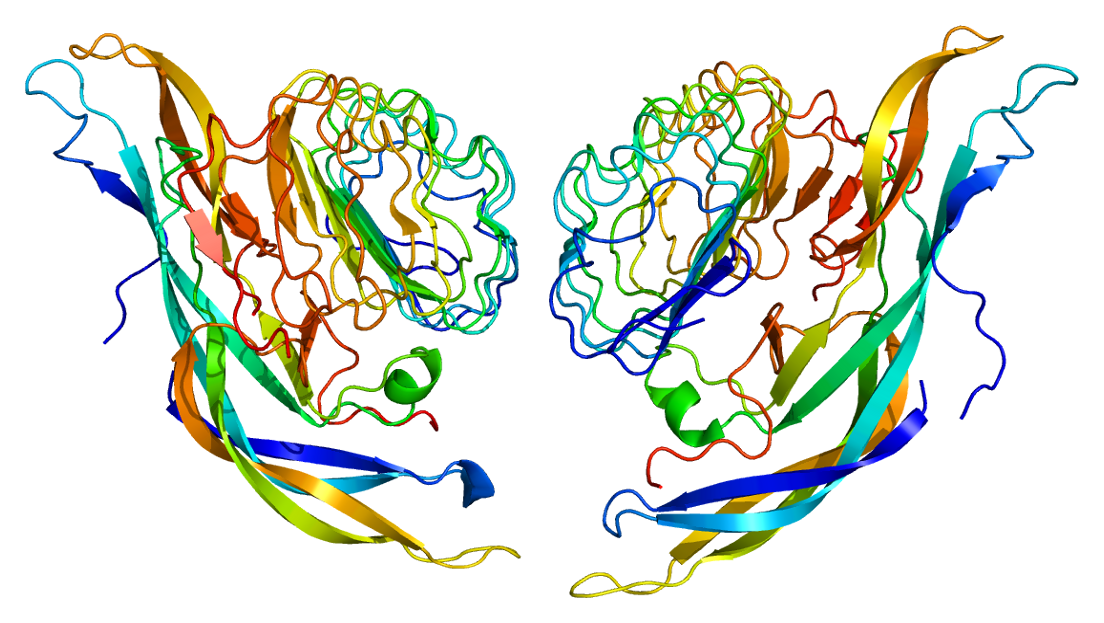
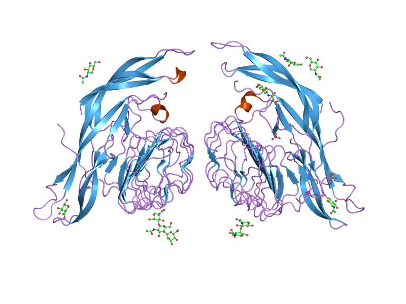
PDB gallery  1xwd: Crystal Structure of Human Follicle Stimulating Hormone Complexed with its Receptor
External links
- "Glycoprotein Hormone Receptors: FSH". IUPHAR Database of Receptors and Ion Channels. International Union of Basic and Clinical Pharmacology. http://www.iuphar-db.org/GPCR/ReceptorDisplayForward?receptorID=2986.
- GRIS: Glycoprotein-hormone Receptor Information System
Cell surface receptor: G protein-coupled receptors Class A:
Rhodopsin likeOtherMetabolites and
signaling moleculesOtherBile acid · Cannabinoid (CB1, CB2, GPR (18, 55, 119)) · EBI2 · Estrogen · Free fatty acid (1, 2, 3, 4) · Lactate · Lysophosphatidic acid (1, 2, 3, 4, 5, 6) · Lysophospholipid (1, 2, 3, 4, 5, 6, 7, 8) · Niacin (1, 2) · Oxoglutarate · PAF · Sphingosine-1-phosphate (1, 2, 3, 4, 5) · SuccinatePeptideOtherAnaphylatoxin (C3a, C5a) · Angiotensin (1, 2) · Apelin · Bombesin (BRS3, GRPR, NMBR) · Bradykinin (B1, B2) · Chemokine · Cholecystokinin (A, B) · Endothelin (A, B) · Formyl peptide (1, 2, 3) · FSH · Galanin (1, 2, 3) · GHB receptor · Gonadotropin-releasing hormone (1, 2) · Ghrelin · Kisspeptin · Luteinizing hormone/choriogonadotropin · MAS (1, 1L, D, E, F, G, X1, X2, X3, X4) · Melanocortin (1, 2, 3, 4, 5) · MCHR (1, 2) · Motilin · Opioid (Delta, Kappa, Mu, Nociceptin & Zeta, but not Sigma) · Orexin (1, 2) · Oxytocin · Prokineticin (1, 2) · Prolactin-releasing peptide · Relaxin (1, 2, 3, 4) · Somatostatin (1, 2, 3, 4, 5) · Tachykinin (1, 2, 3) · Thyrotropin · Thyrotropin-releasing hormone · Urotensin-II · Vasopressin (1A, 1B, 2)MiscellaneousGPR (1, 3, 4, 6, 12, 15, 17, 18, 19, 20, 21, 22, 23, 25, 26, 27, 31, 32, 33, 34, 35, 37, 39, 42, 44, 45, 50, 52, 55, 61, 62, 63, 65, 68, 75, 77, 78, 81, 82, 83, 84, 85, 87, 88, 92, 101, 103, 109A, 109B, 119, 120, 132, 135, 137B, 139, 141, 142, 146, 148, 149, 150, 151, 152, 153, 160, 161, 162, 171, 173, 174, 176, 177, 182, 183)OtherClass B: Secretin like OtherBrain-specific angiogenesis inhibitor (1, 2, 3) · Cadherin (1, 2, 3) · Calcitonin · CALCRL · CD97 · Corticotropin-releasing hormone (1, 2) · EMR (1, 2, 3) · Glucagon (GR, GIPR, GLP1R, GLP2R) · Growth hormone releasing hormone · PACAPR1 · GPR · Latrophilin (1, 2, 3, ELTD1) · Methuselah-like proteins · Parathyroid hormone (1, 2) · Secretin · Vasoactive intestinal peptide (1, 2)Class C: Metabotropic
glutamate / pheromoneOtherClass F:
Frizzled / SmoothenedFrizzledSmoothenedNeuropeptide receptors G protein-coupled receptor OtherOther neuropeptide receptorsAngiotensin · Bradykinin (B1, B2) / Tachykinin (TACR1) · Calcitonin gene-related peptide · Galanin · GPCR neuropeptide (B/W, FF, S, Y) · NeurotensinType I cytokine receptor Enzyme-linked receptor Other Estrogens and progestogens (G03C-D, L02) Progestogens/
progestins
(progesterone)AgonistAndrostene (Drospirenone) • 19-norprogesterone (Nomegestrol • Promegestone • Trimegestone) • 19-nortestosterone (Dienogest)Other/
ungroupedPregnenedione (Gestonorone) • Pregnene (Ethisterone) • Pregnadiene (Medrogestone • Melengestrol) • Norpregnane (Norgestrienone) • Lynestrenol • Norethynodrel • Tibolone • Dydrogesterone • Quingestanolantagonist: MifepristoneAsoprisnil • CDB-4124 • Ulipristal acetateEstrogens AgonistDiosgenin • Estradiol (Ethinylestradiol#/Mestranol • Estradiol 17 beta-cypionate# • Polyestradiol phosphate) • Estrone (Estrone sulfate) • Estriol • Promestriene • Equilenin • EquilinAfimoxifene • Arzoxifene • Bazedoxifene • Cyclofenil • Lasofoxifene • Ormeloxifene • Raloxifene • Tamoxifen • Toremifenepure antagonist: FulvestrantCategories:- Human proteins
- G protein coupled receptors
- Signal transduction
- LRR proteins
Wikimedia Foundation. 2010.