- Cell nucleus
-
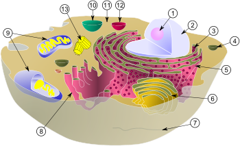 Schematic of typical animal cell, showing subcellular components. Organelles: (1) nucleolus (2) nucleus (3) ribosome (4) vesicle (5) rough endoplasmic reticulum (ER) (6) Golgi apparatus (7) Cytoskeleton (8) smooth ER (9) mitochondria (10) vacuole (11) cytoplasm (12) lysosome (13) centrioles
In cell biology, the nucleus (pl. nuclei; from Latin nucleus or nuculeus, meaning kernel) is a membrane-enclosed organelle found in eukaryotic cells. It contains most of the cell's genetic material, organized as multiple long linear DNA molecules in complex with a large variety of proteins, such as histones, to form chromosomes. The genes within these chromosomes are the cell's nuclear genome. The function of the nucleus is to maintain the integrity of these genes and to control the activities of the cell by regulating gene expression — the nucleus is, therefore, the control center of the cell. The main structures making up the nucleus are the nuclear envelope, a triple cell membrane and membrane that encloses the entire organelle and unifies its contents from the cellular cytoplasm, and the nucleoskeleton (which includes nuclear lamina), a meshwork within the nucleus that adds mechanical support, much like the cytoskeleton, which supports the cell as a whole. Because the nuclear membrane is impermeable to most molecules, nuclear pores are required to allow movement of molecules across the envelope. These pores cross both of the membranes, providing a channel that allows free movement of small molecules and ions. The movement of larger molecules such as proteins is carefully controlled, and requires active transport regulated by carrier proteins. Nuclear transport is crucial to cell function, as movement through the pores is required for both gene expression and chromosomal maintenance. The interior of the nucleus does not contain any membrane-bound subcompartments, its contents are not uniform, and a number of subnuclear bodies exist, made up of unique proteins, RNA molecules, and particular parts of the mitochondria. The best-known of these is the nucleolus, which is mainly involved in the assembly of ribosomes. After being produced in the nucleolus, ribosomes are exported to the cytoplasm where they translate mRNA.
Contents
History
 Oldest known depiction of cells and their nuclei by Antonie van Leeuwenhoek, 1719.
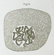 Drawing of a Chironomus salivary gland cell published by Walther Flemming in 1882. The nucleus contains Polytene chromosomes.
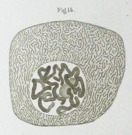
The nucleus was the first organelle to be discovered. The probably oldest preserved drawing dates back to the early microscopist Antonie van Leeuwenhoek (1632 – 1723). He observed a "Lumen", the nucleus, in the red blood cells of salmon.[1] Unlike mammalian red blood cells, those of other vertebrates still possess nuclei. The nucleus was also described by Franz Bauer in 1804[2] and in more detail in 1831 by Scottish botanist Robert Brown in a talk at the Linnean Society of London. Brown was studying orchids under microscope when he observed an opaque area, which he called the areola or nucleus, in the cells of the flower's outer layer.[3] He did not suggest a potential function. In 1838, Matthias Schleiden proposed that the nucleus plays a role in generating cells, thus he introduced the name "Cytoblast" (cell builder). He believed that he had observed new cells assembling around "cytoblasts". Franz Meyen was a strong opponent of this view, having already described cells multiplying by division and believing that many cells would have no nuclei. The idea that cells can be generated de novo, by the "cytoblast" or otherwise, contradicted work by Robert Remak (1852) and Rudolf Virchow (1855) who decisively propagated the new paradigm that cells are generated solely by cells ("Omnis cellula e cellula"). The function of the nucleus remained unclear.[4]
Between 1877 and 1878, Oscar Hedwig published several studies on the fertilization of sea urchin eggs, showing that the nucleus of the sperm enters the oocyte and fuses with its nucleus. This was the first time it was suggested that an individual develops from a (single) nucleated cell. This was in contradiction to Ernst Haeckel's theory that the complete phylogeny of a species would be repeated during embryonic development, including generation of the first nucleated cell from a "Monerula", a structureless mass of primordial mucus ("Urschleim"). Therefore, the necessity of the sperm nucleus for fertilization was discussed for quite some time. However, Hertwig confirmed his observation in other animal groups, e.g., amphibians and molluscs. Eduard Strasburger produced the same results for plants (1884). This paved the way to assign the nucleus an important role in heredity. In 1873, August Weismann postulated the equivalence of the maternal and paternal germ cells for heredity. The function of the nucleus as carrier of genetic information became clear only later, after mitosis was discovered and the Mendelian rules were rediscovered at the beginning of the 20th century; the chromosome theory of heredity was developed.[4]
Structures
The nucleus is the largest cellular organelle in animals.[5] In mammalian cells, the average diameter of the nucleus is approximately 6 micrometers (μm), which occupies about 10% of the total cell volume.[6] The viscous liquid within it is called nucleoplasm, and is similar in composition to the cytosol found outside the nucleus.[7] It appears as a dense, roughly spherical organelle.
Nuclear envelope and pores
Main articles: Nuclear envelope and Nuclear pores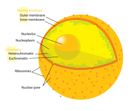 The eukaryotic cell nucleus. Visible in this diagram are the ribosome-studded double membranes of the nuclear envelope, the DNA (complexed as chromatin), and the nucleolus. Within the cell nucleus is a viscous liquid called nucleoplasm, similar to the cytoplasm found outside the nucleus.
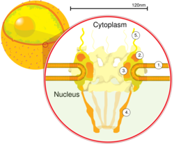 A cross section of a nuclear pore on the surface of the nuclear envelope (1). Other diagram labels show (2) the outer ring, (3) spokes, (4) basket, and (5) filaments.
The outer envelope otherwise known as nuclear membrane consists of two cellular membranes, an inner and an outer membrane, arranged parallel to one another and separated by 10 to 50 nanometers (nm). The nuclear envelope completely encloses the nucleus and separates the cell's genetic material from the surrounding cytoplasm, serving as a barrier to prevent macromolecules from diffusing freely between the nucleoplasm and the cytoplasm.[8] The outer nuclear membrane is continuous with the membrane of the rough endoplasmic reticulum (RER), and is similarly studded with ribosomes.[8] The space between the membranes is called the perinuclear space and is continuous with the RER lumen.
Nuclear pores, which provide aqueous channels through the envelope, are composed of multiple proteins, collectively referred to as nucleoporins. The pores are about 125 million daltons in molecular weight and consist of around 50 (in yeast) to 100 proteins (in vertebrates).[5] The pores are 100 nm in total diameter; however, the gap through which molecules freely diffuse is only about 9 nm wide, due to the presence of regulatory systems within the center of the pore. This size allows the not-free passage of small water-soluble molecules while preventing larger molecules, such as nucleic acids and larger proteins, from inappropriately entering or exiting the nucleus. These large molecules must be actively transported into the nucleus instead. The nucleus of a typical mammalian cell will have about 3000 to 4000 pores throughout its envelope,[9] each of which contains a donut-shaped, eightfold-symmetric ring-shaped structure at a position where the inner and outer membranes fuse.[10] Attached to the ring is a structure called the nuclear basket that extends into the nucleoplasm, and a series of filamentous extensions that reach into the cytoplasm. Both structures serve to mediate binding to nuclear transport proteins.[5]
Most proteins, ribosomal subunits, and some DNAs are transported through the pore complexes in a process mediated by a family of transport factors known as karyopherins. Those karyopherins that mediate movement into the nucleus are also called importins, whereas those that mediate movement out of the nucleus are called exportins. Most karyopherins interact directly with their cargo, although some use adaptor proteins.[11] Steroid hormones such as cortisol and aldosterone, as well as other small lipid-soluble molecules involved in intercellular signaling, can diffuse through the cell membrane and into the cytoplasm, where they bind nuclear receptor proteins that are trafficked into the nucleus. There they serve as transcription factors when bound to their ligand; in the absence of ligand, many such receptors function as histone deacetylases that repress gene expression.[5]
Nuclear lamina
Main article: Nuclear laminaIn animal cells, two networks of intermediate filaments provide the nucleus with mechanical support: The nuclear lamina forms an organized meshwork on the internal face of the envelope, while less organized support is provided on the cytosolic face of the envelope. Both systems provide structural support for the nuclear envelope and anchoring sites for chromosomes and nuclear pores.[6]
The nuclear lamina is composed mostly of lamin proteins. Like all proteins, lamins are synthesized in the cytoplasm and later transported into the nucleus interior, where they are assembled before being incorporated into the existing network of nuclear lamina.[12][13] Lamins found on the cytosolic face of the membrane, such as emerin and nesprin, bind to the cytoskeleton to provide structural support. Lamins are also found inside the nucleoplasm where they form another regular structure, known as the nucleoplasmic veil,[14] that is visible using fluorescence microscopy. The actual function of the veil is not clear, although it is excluded from the nucleolus and is present during interphase.[15] Lamin structures that make up the veil, such as LEM3, bind chromatin and disrupting their structure inhibits transcription of protein-coding genes.[16]
Like the components of other intermediate filaments, the lamin monomer contains an alpha-helical domain used by two monomers to coil around each other, forming a dimer structure called a coiled coil. Two of these dimer structures then join side by side, in an antiparallel arrangement, to form a tetramer called a protofilament. Eight of these protofilaments form a lateral arrangement that is twisted to form a ropelike filament. These filaments can be assembled or disassembled in a dynamic manner, meaning that changes in the length of the filament depend on the competing rates of filament addition and removal.[6]
Mutations in lamin genes leading to defects in filament assembly are known as laminopathies. The most notable laminopathy is the family of diseases known as progeria, which causes the appearance of premature aging in its sufferers. The exact mechanism by which the associated biochemical changes give rise to the aged phenotype is not well understood.[17]
Chromosomes
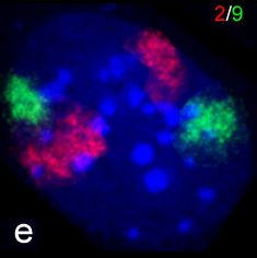
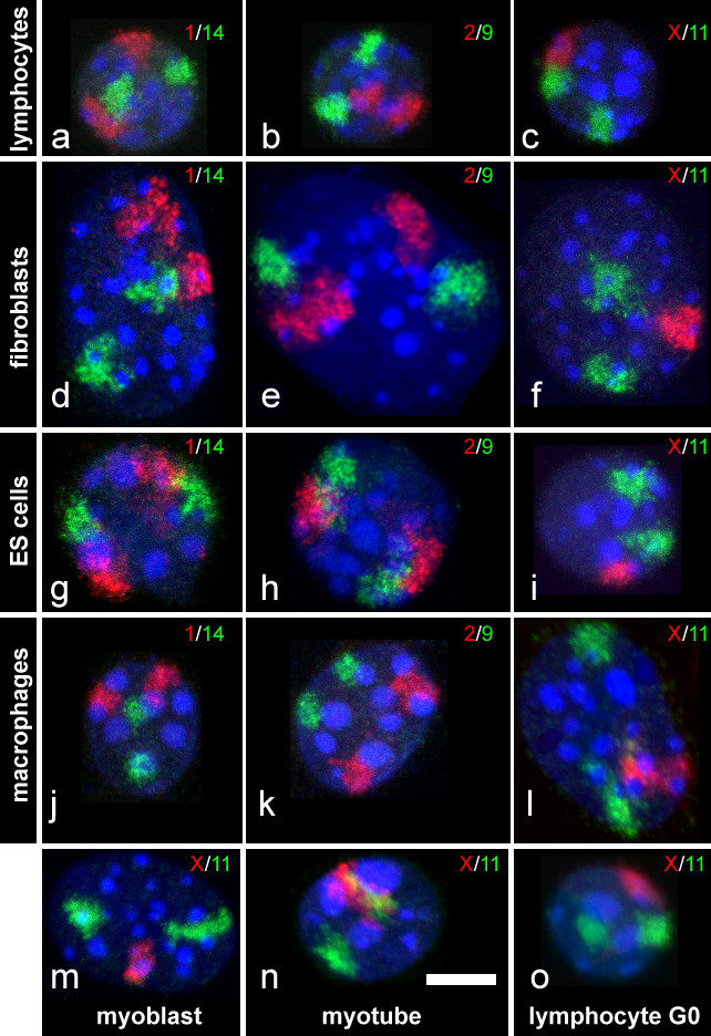
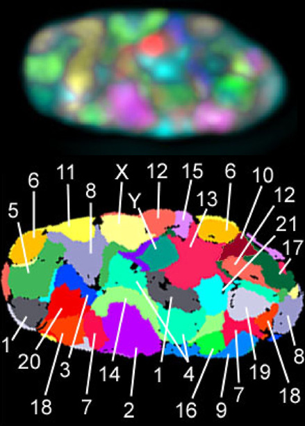
Main article: Chromosome A mouse fibroblast nucleus in which DNA is stained blue. The distinct chromosome territories of chromosome 2 (red) and chromosome 9 (green) are visible stained with fluorescent in situ hybridization.
The cell nucleus contains the majority of the cell's genetic material in the form of multiple linear DNA molecules organized into structures called chromosomes. Each human cell contains 2m of DNA. During most of the cell cycle these are organized in a DNA-protein complex known as chromatin, and during cell division the chromatin can be seen to form the well-defined chromosomes familiar from a karyotype. A small fraction of the cell's genes are located instead in the mitochondria.
There are two types of chromatin. Euchromatin is the less compact DNA form, and contains genes that are frequently expressed by the cell.[18] The other type, heterochromatin, is the more compact form, and contains DNA that are infrequently transcribed. This structure is further categorized into facultative heterochromatin, consisting of genes that are organized as heterochromatin only in certain cell types or at certain stages of development, and constitutive heterochromatin that consists of chromosome structural components such as telomeres and centromeres.[19] During interphase the chromatin organizes itself into discrete individual patches,[20] called chromosome territories.[21] Active genes, which are generally found in the euchromatic region of the chromosome, tend to be located towards the chromosome's territory boundary.[22]
Antibodies to certain types of chromatin organization, in particular, nucleosomes, have been associated with a number of autoimmune diseases, such as systemic lupus erythematosus.[23] These are known as anti-nuclear antibodies (ANA) and have also been observed in concert with multiple sclerosis as part of general immune system dysfunction.[24] As in the case of progeria, the role played by the antibodies in inducing the symptoms of autoimmune diseases is not obvious.
Nucleolus
Main article: Nucleolus An electron micrograph of a cell nucleus, showing the darkly stained nucleolus.
The nucleolus is a discrete densely stained structure found in the nucleus. It is not surrounded by a membrane, and is sometimes called a suborganelle. It forms around tandem repeats of rDNA, DNA coding for ribosomal RNA (rRNA). These regions are called nucleolar organizer regions (NOR). The main roles of the nucleolus are to synthesize rRNA and assemble ribosomes. The structural cohesion of the nucleolus depends on its activity, as ribosomal assembly in the nucleolus results in the transient association of nucleolar components, facilitating further ribosomal assembly, and hence further association. This model is supported by observations that inactivation of rDNA results in intermingling of nucleolar structures.[25]
In the first step of ribosome assembly, a protein called RNA polymerase I transcribes rDNA, which forms a large pre-rRNA precursor. This is cleaved into the subunits 5.8S, 18S, and 28S rRNA.[26] The transcription, post-transcriptional processing, and assembly of rRNA occurs in the nucleolus, aided by small nucleolar RNA (snoRNA) molecules, some of which are derived from spliced introns from messenger RNAs encoding genes related to ribosomal function. The assembled ribosomal subunits are the largest structures passed through the nuclear pores.[5]
When observed under the electron microscope, the nucleolus can be seen to consist of three distinguishable regions: the innermost fibrillar centers (FCs), surrounded by the dense fibrillar component (DFC), which in turn is bordered by the granular component (GC). Transcription of the rDNA occurs either in the FC or at the FC-DFC boundary, and, therefore, when rDNA transcription in the cell is increased, more FCs are detected. Most of the cleavage and modification of rRNAs occurs in the DFC, while the latter steps involving protein assembly onto the ribosomal subunits occur in the GC.[26]
Other subnuclear bodies
Subnuclear structure sizes Structure name Structure diameter Cajal bodies 0.2–2.0 µm [27] PIKA 5 µm [28] PML bodies 0.2–1.0 µm [29] Paraspeckles 0.2–1.0 µm [30] Speckles 20–25 nm [28] Besides the nucleolus, the nucleus contains a number of other non-membrane-delineated bodies. These include Cajal bodies, Gemini of coiled bodies, polymorphic interphase karyosomal association (PIKA), promyelocytic leukaemia (PML) bodies, paraspeckles, and splicing speckles. Although little is known about a number of these domains, they are significant in that they show that the nucleoplasm is not uniform mixture, but rather contains organized functional subdomains.[29]
Other subnuclear structures appear as part of abnormal disease processes. For example, the presence of small intranuclear rods has been reported in some cases of nemaline myopathy. This condition typically results from mutations in actin, and the rods themselves consist of mutant actin as well as other cytoskeletal proteins.[31]
Cajal bodies and gems
A nucleus typically contains between 1 and 10 compact structures called Cajal bodies or coiled bodies (CB), whose diameter measures between 0.2 µm and 2.0 µm depending on the cell type and species.[27] When seen under an electron microscope, they resemble balls of tangled thread[28] and are dense foci of distribution for the protein coilin.[32] CBs are involved in a number of different roles relating to RNA processing, specifically small nucleolar RNA (snoRNA) and small nuclear RNA (snRNA) maturation, and histone mRNA modification.[27]
Similar to Cajal bodies are Gemini of coiled bodies, or gems, whose name is derived from the Gemini constellation in reference to their close "twin" relationship with CBs. Gems are similar in size and shape to CBs, and in fact are virtually indistinguishable under the microscope.[32] Unlike CBs, gems do not contain small nuclear ribonucleoproteins (snRNPs), but do contain a protein called survivor of motor neurons (SMN) whose function relates to snRNP biogenesis. Gems are believed to assist CBs in snRNP biogenesis,[33] though it has also been suggested from microscopy evidence that CBs and gems are different manifestations of the same structure.[32]
RAFA and PTF domains
RAFA domains, or polymorphic interphase karyosomal associations, were first described in microscopy studies in 1991. Their function was and remains unclear, though they were not thought to be associated with active DNA replication, transcription, or RNA processing.[34] They have been found to often associate with discrete domains defined by dense localization of the transcription factor PTF, which promotes transcription of snRNA.[35]
PML bodies
Promyelocytic leukaemia bodies (PML bodies) are spherical bodies found scattered throughout the nucleoplasm, measuring around 0.2–1.0 µm. They are known by a number of other names, including nuclear domain 10 (ND10), Kremer bodies, and PML oncogenic domains. They are often seen in the nucleus in association with Cajal bodies and cleavage bodies. It has been suggested that they play a role in regulating transcription.[29]
Paraspeckles
Main article: ParaspeckleDiscovered by Fox et al. in 2002, paraspeckles are irregularly shaped compartments in the nucleus' interchromatin space.[36] First documented in HeLa cells, where there are generally 10–30 per nucleus,[37] paraspeckles are now known to also exist in all human primary cells, transformed cell lines, and tissue sections.[38] Their name is derived from their distribution in the nucleus; the "para" is short for parallel and the "speckles" refers to the splicing speckles to which they are always in close proximity.[37]
Paraspeckles are dynamic structures that are altered in response to changes in cellular metabolic activity. They are transcription dependent[36] and in the absence of RNA Pol II transcription, the paraspeckle disappears and all of its associated protein components (PSP1, p54nrb, PSP2, CFI(m)68, and PSF) form a crescent shaped perinucleolar cap in the nucleolus. This phenomenon is demonstrated during the cell cycle. In the cell cycle, paraspeckles are present during interphase and during all of mitosis except for telophase. During telophase, when the two daughter nuclei are formed, there is no RNA Pol II transcription so the protein components instead form a perinucleolar cap.[38]
Splicing speckles
Speckles are subnuclear structures that are enriched in pre-messenger RNA splicing factors and are located in the interchromatin regions of the nucleoplasm of mammalian cells. At the fluorescence-microscope level they appear as irregular, punctate structures, which vary in size and shape, and when examined by electron microscopy they are seen as clusters of interchromatin granules. Speckles are dynamic structures, and both their protein and RNA-protein components can cycle continuously between speckles and other nuclear locations, including active transcription sites. Studies on the composition, structure and behaviour of speckles have provided a model for understanding the functional compartmentalization of the nucleus and the organization of the gene-expression machinery.[39]
Sometimes referred to as interchromatin granule clusters or as splicing-factor compartments, speckles are rich in splicing snRNPs[40][41] and other splicing proteins necessary for pre-mRNA processing.[42] Because of a cell's changing requirements, the composition and location of these bodies changes according to mRNA transcription and regulation via phosphorylation of specific proteins.[43]
Function
The main function of the cell nucleus is to control gene expression and mediate the replication of DNA during the cell cycle. The nucleus provides a site for genetic transcription that is segregated from the location of translation in the cytoplasm, allowing levels of gene regulation that are not available to prokaryotes.
Cell compartmentalization
The nuclear envelope allows the nucleus to control its contents, and separate them from the rest of the cytoplasm where necessary. This is important for controlling processes on either side of the nuclear membrane. In some cases where a cytoplasmic process needs to be restricted, a key participant is removed to the nucleus, where it interacts with transcription factors to downregulate the production of certain enzymes in the pathway. This regulatory mechanism occurs in the case of glycolysis, a cellular pathway for breaking down glucose to produce energy. Hexokinase is an enzyme responsible for the first the step of glycolysis, forming glucose-6-phosphate from glucose. At high concentrations of fructose-6-phosphate, a molecule made later from glucose-6-phosphate, a regulator protein removes hexokinase to the nucleus,[44] where it forms a transcriptional repressor complex with nuclear proteins to reduce the expression of genes involved in glycolysis.[45]
In order to control which genes are being transcribed, the cell separates some transcription factor proteins responsible for regulating gene expression from physical access to the DNA until they are activated by other signaling pathways. This prevents even low levels of inappropriate gene expression. For example, in the case of NF-κB-controlled genes, which are involved in most inflammatory responses, transcription is induced in response to a signal pathway such as that initiated by the signaling molecule TNF-α, binds to a cell membrane receptor, resulting in the recruitment of signalling proteins, and eventually activating the transcription factor NF-κB. A nuclear localisation signal on the NF-κB protein allows it to be transported through the nuclear pore and into the nucleus, where it stimulates the transcription of the target genes.[6]
The compartmentalization allows the cell to prevent translation of unspliced mRNA.[46] Eukaryotic mRNA contains introns that must be removed before being translated to produce functional proteins. The splicing is done inside the nucleus before the mRNA can be accessed by ribosomes for translation. Without the nucleus, ribosomes would translate newly transcribed (unprocessed) mRNA, resulting in misformed and nonfunctional proteins.
Gene expression
Main article: Gene expression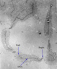 A micrograph of ongoing gene transcription of ribosomal RNA illustrating the growing primary transcripts. "Begin" indicates the 3' end of the DNA, where new RNA synthesis begins; "end" indicates the 5' end, where the primary transcripts are almost complete.
Gene expression first involves transcription, in which DNA is used as a template to produce RNA. In the case of genes encoding proteins, that RNA produced from this process is messenger RNA (mRNA), which then needs to be translated by ribosomes to form a protein. As ribosomes are located outside the nucleus, mRNA produced needs to be exported.[47]
Since the nucleus is the site of transcription, it also contains a variety of proteins that either directly mediate transcription or are involved in regulating the process. These proteins include helicases, which unwind the double-stranded DNA molecule to facilitate access to it, RNA polymerases, which synthesize the growing RNA molecule, topoisomerases, which change the amount of supercoiling in DNA, helping it wind and unwind, as well as a large variety of transcription factors that regulate expression.[48]
Processing of pre-mRNA
Main article: Post-transcriptional modificationNewly synthesized mRNA molecules are known as primary transcripts or pre-mRNA. They must undergo post-transcriptional modification in the nucleus before being exported to the cytoplasm; mRNA that appears in the cytoplasm without these modifications is degraded rather than used for protein translation. The three main modifications are 5' capping, 3' polyadenylation, and RNA splicing. While in the nucleus, pre-mRNA is associated with a variety of proteins in complexes known as heterogeneous ribonucleoprotein particles (hnRNPs). Addition of the 5' cap occurs co-transcriptionally and is the first step in post-transcriptional modification. The 3' poly-adenine tail is only added after transcription is complete.
RNA splicing, carried out by a complex called the spliceosome, is the process by which introns, or regions of DNA that do not code for protein, are removed from the pre-mRNA and the remaining exons connected to re-form a single continuous molecule. This process normally occurs after 5' capping and 3' polyadenylation but can begin before synthesis is complete in transcripts with many exons.[5] Many pre-mRNAs, including those encoding antibodies, can be spliced in multiple ways to produce different mature mRNAs that encode different protein sequences. This process is known as alternative splicing, and allows production of a large variety of proteins from a limited amount of DNA.
Dynamics and regulation
Nuclear transport
Main article: Nuclear transport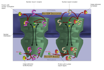 Macromolecules, such as RNA and proteins, are actively transported across the nuclear membrane in a process called the Ran-GTP nuclear transport cycle.
The entry and exit of large molecules from the nucleus is tightly controlled by the nuclear pore complexes. Although small molecules can enter the nucleus without regulation,[49] macromolecules such as RNA and proteins require association karyopherins called importins to enter the nucleus and exportins to exit. "Cargo" proteins that must be translocated from the cytoplasm to the nucleus contain short amino acid sequences known as nuclear localization signals, which are bound by importins, while those transported from the nucleus to the cytoplasm carry nuclear export signals bound by exportins. The ability of importins and exportins to transport their cargo is regulated by GTPases, enzymes that hydrolyze the molecule guanosine triphosphate to release energy. The key GTPase in nuclear transport is Ran, which can bind either GTP or GDP (guanosine diphosphate), depending on whether it is located in the nucleus or the cytoplasm. Whereas importins depend on RanGTP to dissociate from their cargo, exportins require RanGTP in order to bind to their cargo.[11]
Nuclear import depends on the importin binding its cargo in the cytoplasm and carrying it through the nuclear pore into the nucleus. Inside the nucleus, RanGTP acts to separate the cargo from the importin, allowing the importin to exit the nucleus and be reused. Nuclear export is similar, as the exportin binds the cargo inside the nucleus in a process facilitated by RanGTP, exits through the nuclear pore, and separates from its cargo in the cytoplasm.
Specialized export proteins exist for translocation of mature mRNA and tRNA to the cytoplasm after post-transcriptional modification is complete. This quality-control mechanism is important due to the these molecules' central role in protein translation; mis-expression of a protein due to incomplete excision of exons or mis-incorporation of amino acids could have negative consequences for the cell; thus, incompletely modified RNA that reaches the cytoplasm is degraded rather than used in translation.[5]
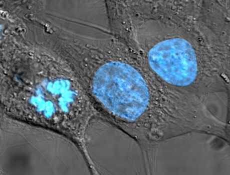
Assembly and disassembly
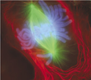
 An image of a newt lung cell stained with fluorescent dyes during metaphase. The mitotic spindle can be seen, stained green, attached to the two sets of chromosomes, stained light blue. All chromosomes but one are already at the metaphase plate.
During its lifetime, a nucleus may be broken down, either in the process of cell division or as a consequence of apoptosis, a regulated form of cell death. During these events, the structural components of the nucleus — the envelope and lamina — can be systematically degraded. In most cells, the disassembly of the nuclear envelope marks the end of the prophase of mitosis. However, this disassembly of the nucleus is not a universal feature of mitosis and does not occur in all cells. Some unicellular eukaryotes (e.g., yeasts) undergo so-called closed mitosis, in which the nuclear envelope remains intact. In closed mitosis, the daughter chromosomes migrate to opposite poles of the nucleus, which then divides in two. The cells of higher eukaryotes, however, usually undergo open mitosis, which is characterized by breakdown of the nuclear envelope. The daughter chromosomes then migrate to opposite poles of the mitotic spindle, and new nuclei reassemble around them
At a certain point during the cell cycle in open mitosis, the cell divides to form two cells. In order for this process to be possible, each of the new daughter cells must have a full set of genes, a process requiring replication of the chromosomes as well as segregation of the separate sets. This occurs by the replicated chromosomes, the sister chromatids, attaching to microtubules, which in turn are attached to different centrosomes. The sister chromatids can then be pulled to separate locations in the cell. In many cells, the centrosome is located in the cytoplasm, outside the nucleus; the microtubules would be unable to attach to the chromatids in the presence of the nuclear envelope.[50] Therefore the early stages in the cell cycle, beginning in prophase and until around prometaphase, the nuclear membrane is dismantled.[14] Likewise, during the same period, the nuclear lamina is also disassembled, a process regulated by phosphorylation of the lamins by protein kinases such as the CDC2 protein kinase.[51] Towards the end of the cell cycle, the nuclear membrane is reformed, and around the same time, the nuclear lamina are reassembled by dephosphorylating the lamins.[51]
However, in dinoflagellates, the nuclear envelope remains intact, the centrosomes are located in the cytoplasm, and the microtubules come in contact with chromosomes, whose centromeric regions are incorporated into the nuclear envelope (the so-called closed mitosis with extranuclear spindle). In many other protists (e.g., ciliates, sporozoans) and fungi, the centrosomes are intranuclear, and their nuclear envelope also does not disassemle during cell division.
Apoptosis is a controlled process in which the cell's structural components are destroyed, resulting in death of the cell. Changes associated with apoptosis directly affect the nucleus and its contents, for example, in the condensation of chromatin and the disintegration of the nuclear envelope and lamina. The destruction of the lamin networks is controlled by specialized apoptotic proteases called caspases, which cleave the lamin proteins and, thus, degrade the nucleus' structural integrity. Lamin cleavage is sometimes used as a laboratory indicator of caspase activity in assays for early apoptotic activity.[14] Cells that express mutant caspase-resistant lamins are deficient in nuclear changes related to apoptosis, suggesting that lamins play a role in initiating the events that lead to apoptotic degradation of the nucleus.[14] Inhibition of lamin assembly itself is an inducer of apoptosis.[52]
The nuclear envelope acts as a barrier that prevents both DNA and RNA viruses from entering the nucleus. Some viruses require access to proteins inside the nucleus in order to replicate and/or assemble. DNA viruses, such as herpesvirus replicate and assemble in the cell nucleus, and exit by budding through the inner nuclear membrane. This process is accompanied by disassembly of the lamina on the nuclear face of the inner membrane.[14]
Anucleated and polynucleated cells
 Human red blood cells, like those of other mammals, lack nuclei. This occurs as a normal part of the cells' development.
Although most cells have a single nucleus, some eukaryotic cell types have no nucleus, and others have many nuclei. This can be a normal process, as in the maturation of mammalian red blood cells, or a result of faulty cell division.
Anucleated cells contain no nucleus and are, therefore, incapable of dividing to produce daughter cells. The best-known anucleated cell is the mammalian red blood cell, or erythrocyte, which also lacks other organelles such as mitochondria, and serves primarily as a transport vessel to ferry oxygen from the lungs to the body's tissues. Erythrocytes mature through erythropoiesis in the bone marrow, where they lose their nuclei, organelles, and ribosomes. The nucleus is expelled during the process of differentiation from an erythroblast to a reticulocyte, which is the immediate precursor of the mature erythrocyte.[53] The presence of mutagens may induce the release of some immature "micronucleated" erythrocytes into the bloodstream.[54][55] Anucleated cells can also arise from flawed cell division in which one daughter lacks a nucleus and the other has two nuclei.
Polynucleated cells contain multiple nuclei. Most Acantharean species of protozoa[56] and some fungi in mycorrhizae[57] have naturally polynucleated cells. Other examples include the intestinal parasites in the genus Giardia, which have two nuclei per cell.[58] In humans, skeletal muscle cells, called myocytes, become polynucleated during development; the resulting arrangement of nuclei near the periphery of the cells allows maximal intracellular space for myofibrils.[5] Multinucleated and Binucleated cells can also be abnormal in humans; for example, cells arising from the fusion of monocytes and macrophages, known as giant multinucleated cells, sometimes accompany inflammation[59] and are also implicated in tumor formation.[60]
Evolution
As the major defining characteristic of the eukaryotic cell, the nucleus' evolutionary origin has been the subject of much speculation. Four major theories have been proposed to explain the existence of the nucleus, although none have yet earned widespread support.[61]
The theory known as the "syntrophic model" proposes that a symbiotic relationship between the archaea and bacteria created the nucleus-containing eukaryotic cell. (Organisms of the Archaea domain have no cell nucleus.[62]) It is hypothesized that the symbiosis originated when ancient archaea, similar to modern methanogenic archaea, invaded and lived within bacteria similar to modern myxobacteria, eventually forming the early nucleus. This theory is analogous to the accepted theory for the origin of eukaryotic mitochondria and chloroplasts, which are thought to have developed from a similar endosymbiotic relationship between proto-eukaryotes and aerobic bacteria.[63] The archaeal origin of the nucleus is supported by observations that archaea and eukarya have similar genes for certain proteins, including histones. Observations that myxobacteria are motile, can form multicellular complexes, and possess kinases and G proteins similar to eukarya, support a bacterial origin for the eukaryotic cell.[64]
A second model proposes that proto-eukaryotic cells evolved from bacteria without an endosymbiotic stage. This model is based on the existence of modern planctomycetes bacteria that possess a nuclear structure with primitive pores and other compartmentalized membrane structures.[65] A similar proposal states that a eukaryote-like cell, the chronocyte, evolved first and phagocytosed archaea and bacteria to generate the nucleus and the eukaryotic cell.[66]
The most controversial model, known as viral eukaryogenesis, posits that the membrane-bound nucleus, along with other eukaryotic features, originated from the infection of a prokaryote by a virus. The suggestion is based on similarities between eukaryotes and viruses such as linear DNA strands, mRNA capping, and tight binding to proteins (analogizing histones to viral envelopes). One version of the proposal suggests that the nucleus evolved in concert with phagocytosis to form an early cellular "predator".[67] Another variant proposes that eukaryotes originated from early archaea infected by poxviruses, on the basis of observed similarity between the DNA polymerases in modern poxviruses and eukaryotes.[68][69] It has been suggested that the unresolved question of the evolution of sex could be related to the viral eukaryogenesis hypothesis.[70]
A very recent proposal suggests that traditional variants of the endosymbiont theory are insufficiently powerful to explain the origin of the eukaryotic nucleus. This model, termed the exomembrane hypothesis, suggests that the nucleus instead originated from a single ancestral cell that evolved a second exterior cell membrane; the interior membrane enclosing the original cell then became the nuclear membrane and evolved increasingly elaborate pore structures for passage of internally synthesized cellular components such as ribosomal subunits.[71]
References
- ^ Leeuwenhoek, A. van: Opera Omnia, seu Arcana Naturae ope exactissimorum Microscopiorum detecta, experimentis variis comprobata, Epistolis ad varios illustres viros. J. Arnold et Delphis, A. Beman, Lugdinum Batavorum 1719–1730. Cited after: Dieter Gerlach, Geschichte der Mikroskopie. Verlag Harry Deutsch, Frankfurt am Main, Germany, 2009. ISBN 978-3-8171-1781-9.
- ^ Harris, H (1999). The Birth of the Cell. New Haven: Yale University Press. ISBN 0-300-07384-4.
- ^ Brown, Robert (1866). "On the Organs and Mode of Fecundation of Orchidex and Asclepiadea". Miscellaneous Botanical Works I: 511–514.
- ^ a b Cremer, Thomas (1985). Von der Zellenlehre zur Chromosomentheorie. Berlin, Heidelberg, New York, Tokyo: Springer Verlag. ISBN 3-540-13987-7. Online Version here
- ^ a b c d e f g h Lodish, H; Berk A, Matsudaira P, Kaiser CA, Krieger M, Scott MP, Zipursky SL, Darnell J. (2004). Molecular Cell Biology (5th ed.). New York: WH Freeman. ISBN 0-7167-2672-6.
- ^ a b c d Bruce Alberts, Alexander Johnson, Julian Lewis, Martin Raff, Keith Roberts, Peter Walter, ed (2002). Molecular Biology of the Cell, Chapter 4, pages 191–234 (4th ed.). Garland Science.
- ^ Clegg JS (February 1984). "Properties and metabolism of the aqueous cytoplasm and its boundaries". Am. J. Physiol. 246 (2 Pt 2): R133–51. PMID 6364846. http://ajpregu.physiology.org/cgi/pmidlookup?view=reprint&pmid=6364846.
- ^ a b Paine P, Moore L, Horowitz S (1975). "Nuclear envelope permeability". Nature 254 (5496): 109–114. doi:10.1038/254109a0. PMID 1117994.
- ^ Rodney Rhoades, Richard Pflanzer, ed (1996). "Ch3". Human Physiology (3rd ed.). Saunders College Publishing.
- ^ Shulga N, Mosammaparast N, Wozniak R, Goldfarb D (2000). "Yeast nucleoporins involved in passive nuclear envelope permeability". J Cell Biol 149 (5): 1027–1038. doi:10.1083/jcb.149.5.1027. PMID 10831607.
- ^ a b Pemberton L, Paschal B (2005). "Mechanisms of receptor-mediated nuclear import and nuclear export". Traffic 6 (3): 187–198. doi:10.1111/j.1600-0854.2005.00270.x. PMID 15702987.
- ^ Stuurman N, Heins S, Aebi U (1998). "Nuclear lamins: their structure, assembly, and interactions". J Struct Biol 122 (1–2): 42–66. doi:10.1006/jsbi.1998.3987. PMID 9724605.
- ^ Goldman A, Moir R, Montag-Lowy M, Stewart M, Goldman R (1992). "Pathway of incorporation of microinjected lamin A into the nuclear envelope". J Cell Biol 119 (4): 725–735. doi:10.1083/jcb.119.4.725. PMID 1429833.
- ^ a b c d e Goldman R, Gruenbaum Y, Moir R, Shumaker D, Spann T (2002). "Nuclear lamins: building blocks of nuclear architecture". Genes Dev 16 (5): 533–547. doi:10.1101/gad.960502. PMID 11877373. http://www.genesdev.org/cgi/content/full/16/5/533.
- ^ Moir RD, Yoona M, Khuona S, Goldman RD. (2000). "Nuclear Lamins A and B1: Different Pathways of Assembly during Nuclear Envelope Formation in Living Cells". Journal of Cell Biology 151 (6): 1155–1168. doi:10.1083/jcb.151.6.1155. PMID 11121432.
- ^ Spann TP, Goldman AE, Wang C, Huang S, Goldman RD. (2002). "Alteration of nuclear lamin organization inhibits RNA polymerase II–dependent transcription". Journal of Cell Biology 156 (4): 603–608. doi:10.1083/jcb.200112047. PMID 11854306.
- ^ Mounkes LC, Stewart CL (2004). "Aging and nuclear organization: lamins and progeria". Current Opinion in Cell Biology 16 (3): 322–327. doi:10.1016/j.ceb.2004.03.009. PMID 15145358.
- ^ Ehrenhofer-Murray A (2004). "Chromatin dynamics at DNA replication, transcription and repair". Eur J Biochem 271 (12): 2335–2349. doi:10.1111/j.1432-1033.2004.04162.x. PMID 15182349.
- ^ Grigoryev S, Bulynko Y, Popova E (2006). "The end adjusts the means: heterochromatin remodelling during terminal cell differentiation". Chromosome Res 14 (1): 53–69. doi:10.1007/s10577-005-1021-6. PMID 16506096.
- ^ Schardin, Margit; Cremer, T; Hager, HD; Lang, M (December 1985). "Specific staining of human chromosomes in Chinese hamster x man hybrid cell lines demonstrates interphase chromosome territories". Human Genetics (Springer Berlin / Heidelberg) 71 (4): 281–287. doi:10.1007/BF00388452. PMID 2416668. http://www.springerlink.com/content/lv101t8w17306071/.
- ^ Lamond, Angus I.; William C. Earnshaw (1998-04-24). "Structure and Function in the Nucleus". Science 280 (5363): 547–553. doi:10.1126/science.280.5363.547. PMID 9554838.
- ^ Kurz, A; Lampel, S; Nickolenko, JE; Bradl, J; Benner, A; Zirbel, RM; Cremer, T; Lichter, P (1996). "Active and inactive genes localize preferentially in the periphery of chromosome territories". The Journal of Cell Biology (The Rockefeller University Press) 135 (5): 1195–1205. doi:10.1083/jcb.135.5.1195. PMC 2121085. PMID 8947544. http://intl.jcb.org/cgi/content/abstract/135/5/1195.
- ^ NF Rothfield, BD Stollar (1967). "The Relation of Immunoglobulin Class, Pattern of Antinuclear Antibody, and Complement-Fixing Antibodies to DNA in Sera from Patients with Systemic Lupus Erythematosus". J Clin Invest 46 (11): 1785–1794. doi:10.1172/JCI105669. PMC 292929. PMID 4168731. http://www.pubmedcentral.nih.gov/articlerender.fcgi?tool=pmcentrez&artid=292929.
- ^ S Barned, AD Goodman, DH Mattson (1995). "Frequency of anti-nuclear antibodies in multiple sclerosis". Neurology 45 (2): 384–385. PMID 7854544.
- ^ Hernandez-Verdun, Daniele (2006). "Nucleolus: from structure to dynamics". Histochem. Cell. Biol 125 (1-2): 127–137. doi:10.1007/s00418-005-0046-4. PMID 16328431.
- ^ a b Lamond, Angus I.; Judith E. Sleeman. "Nuclear substructure and dynamics". current biology 13 (21): R825–828. doi:10.1016/j.cub.2003.10.012. PMID 14588256.
- ^ a b c Cioce M, Lamond A (2005). "Cajal bodies: a long history of discovery". Annu Rev Cell Dev Biol 21: 105–131. doi:10.1146/annurev.cellbio.20.010403.103738. PMID 16212489.
- ^ a b c Pollard, Thomas D.; William C. Earnshaw (2004). Cell Biology. Philadelphia: Saunders. ISBN 0-7216-3360-9.
- ^ a b c Dundr, Miroslav; Tom Misteli (2001). "Functional architecture in the cell nucleus". Biochem. J. (356): 297–310. PMID 11368755.
- ^ Fox, Archa (2007-03-07). Paraspeckle Size. Interview with R. Sundby. E-mail Correspondence.
- ^ Goebel, H.H.; I Warlow (January 1997). "Nemaline myopathy with intranuclear rods—intranuclear rod myopathy". Neuromuscular Disorders 7 (1): 13–19. doi:10.1016/S0960-8966(96)00404-X. PMID 9132135.
- ^ a b c Matera AG, Frey MA. (1998). "Coiled Bodies and Gems: Janus or Gemini?". American Journal of Human Genetics 63 (2): 317–321. doi:10.1086/301992. PMID 9683623.
- ^ Matera, A. Gregory (1998). "Of Coiled Bodies, Gems, and Salmon". Journal of Cellular Biochemistry (70): 181–192. PMID 9671224.
- ^ Saunders WS, Cooke CA, Earnshaw WC (1991). "Compartmentalization within the nucleus: discovery of a novel subnuclear region.". Journal of Cellular Biology 115 (4): 919–931. doi:10.1083/jcb.115.4.919. PMID 1955462
- ^ Pombo A, Cuello P, Schul W, Yoon J, Roeder R, Cook P, Murphy S (1998). "Regional and temporal specialization in the nucleus: a transcriptionally active nuclear domain rich in PTF, Oct1 and PIKA antigens associates with specific chromosomes early in the cell cycle". EMBO J 17 (6): 1768–1778. doi:10.1093/emboj/17.6.1768. PMID 9501098.
- ^ a b Fox, Archa; Lam, YW; Leung, AK; Lyon, CE; Andersen, J; Mann, M; Lamond, AI (2002). "Paraspeckles:A Novel Nuclear Domain". Current Biology 12 (1): 13–25. doi:10.1016/S0960-9822(01)00632-7. PMID 11790299. http://www.current-biology.com/content/article/abstract?uid=PIIS0960982201006327.
- ^ a b Fox, Archa; Wendy Bickmore (2004). "Nuclear Compartments: Paraspeckles". Nuclear Protein Database. Archived from the original on May 2, 2006. http://web.archive.org/web/20060502134554/http://npd.hgu.mrc.ac.uk/compartments/paraspeckles.html. Retrieved 2007-03-06.
- ^ a b Fox, A. et al. (2005). "P54nrb Forms a Heterodimer with PSP1 That Localizes to Paraspeckles in an RNA-dependent Manner". Molecular Biology of the Cell 16 (11): 5304–5315. doi:10.1091/mbc.E05-06-0587. PMC 1266428. PMID 16148043. http://www.molbiolcell.org/cgi/reprint/16/11/5304.
- ^ Lamond AI, Spector DL (August 2003). "Nuclear speckles: a model for nuclear organelles". Nat. Rev. Mol. Cell Biol. 4 (8): 605–12. doi:10.1038/nrm1172. PMID 12923522.
- ^ Tripathi K, Parnaik VK (September 2008). "Differential dynamics of splicing factor SC35 during the cell cycle" (PDF). J. Biosci. 33 (3): 345–54. PMID 19005234. http://www.ias.ac.in/jbiosci/sep2008/345.pdf.
- ^ Tripathi, K; Parnaik, VK (2008). "Differential dynamics of splicing factor SC35 during the cell cycle". Journal of biosciences 33 (3): 345–54. PMID 19005234.
- ^ Lamond AI, Spector DL (August 2003). "Nuclear speckles: a model for nuclear organelles". Nat. Rev. Mol. Cell Biol. 4 (8): 605–12. doi:10.1038/nrm1172. PMID 12923522.
- ^ Handwerger, Korie E.; Joseph G. Gall (January 2006). "Subnuclear organelles: new insights into form and function". TRENDS in Cell Biology 16 (1): 19–26. doi:10.1016/j.tcb.2005.11.005. PMID 16325406.
- ^ Lehninger, Albert L.; David L. Nelson, Michael M. Cox. (2000). Lehninger principles of biochemistry (3rd ed.). New York: Worth Publishers. ISBN 1-57259-931-6.
- ^ Moreno F, Ahuatzi D, Riera A, Palomino CA, Herrero P. (2005). "Glucose sensing through the Hxk2-dependent signalling pathway.". Biochem Soc Trans 33 (1): 265–268. doi:10.1042/BST0330265. PMID 15667322. PMID 15667322
- ^ Görlich, Dirk; Ulrike Kutay (1999). "Transport between the cell nucleus and the cytoplasm". Ann. Rev. Cell Dev. Biol. 15 (1): 607–660. doi:10.1146/annurev.cellbio.15.1.607. PMID 10611974.
- ^ Nierhaus, Knud H.; Daniel N. Wilson (2004). Protein Synthesis and Ribosome Structure: Translating the Genome. Wiley-VCH. ISBN 3-527-30638-2.
- ^ Nicolini, Claudio A. (1997). Genome Structure and Function: From Chromosomes Characterization to Genes Technology. Springer. ISBN 0-7923-4565-7.
- ^ Watson, JD; Baker TA, Bell SP, Gann A, Levine M, Losick R. (2004). "Ch9–10". Molecular Biology of the Gene (5th ed.). Peason Benjamin Cummings; CSHL Press.. ISBN 0-8053-9603-9.
- ^ Lippincott-Schwartz, Jennifer (2002-03-07). "Cell biology: Ripping up the nuclear envelope". Nature 416 (6876): 31–32. doi:10.1038/416031a. PMID 11882878.
- ^ a b Boulikas T (1995). "Phosphorylation of transcription factors and control of the cell cycle". Crit Rev Eukaryot Gene Expr 5 (1): 1–77. PMID 7549180.
- ^ Steen R, Collas P (2001). "Mistargeting of B-type lamins at the end of mitosis: implications on cell survival and regulation of lamins A/C expression". J Cell Biol 153 (3): 621–626. doi:10.1083/jcb.153.3.621. PMID 11331311.
- ^ Skutelsky, E.; Danon D. (June 1970). "Comparative study of nuclear expulsion from the late erythroblast and cytokinesis". J Cell Biol (60(3)): 625–635. doi:10.1016/0014-4827(70)90536-7. PMID 5422968.
- ^ Torous, DK; Dertinger SD, Hall NE, Tometsko CR. (2000). "Enumeration of micronucleated reticulocytes in rat peripheral blood: a flow cytometric study". Mutat Res (465(1–2)): 91–99. PMID 10708974.
- ^ Hutter, KJ; Stohr M. (1982). "Rapid detection of mutagen induced micronucleated erythrocytes by flow cytometry". Histochemistry 75 (3): 353–362. PMID 7141888.
- ^ Zettler, LA; Sogin ML, Caron DA (1997). "Phylogenetic relationships between the Acantharea and the Polycystinea: A molecular perspective on Haeckel's Radiolaria". Proc Natl Acad Sci USA 94 (21): 11411–11416. doi:10.1073/pnas.94.21.11411. PMC 23483. PMID 9326623. http://www.pubmedcentral.nih.gov/articlerender.fcgi?tool=pmcentrez&artid=23483.
- ^ Horton, TR (2006). "The number of nuclei in basidiospores of 63 species of ectomycorrhizal Homobasidiomycetes". Mycologia 98 (2): 233–238. doi:10.3852/mycologia.98.2.233. PMID 16894968.
- ^ Adam RD (December 1991). "The biology of Giardia spp". Microbiol. Rev. 55 (4): 706–32. PMC 372844. PMID 1779932. http://mmbr.asm.org/cgi/pmidlookup?view=long&pmid=1779932.
- ^ McInnes, A; Rennick DM (1988). "Interleukin 4 induces cultured monocytes/macrophages to form giant multinucleated cells". J Exp Med (167): 598–611. doi:10.1084/jem.167.2.598. PMID 3258008.
- ^ Goldring, SR; Roelke MS, Petrison KK, Bhan AK (1987). "Human giant cell tumors of bone identification and characterization of cell types". J Clin Invest 79 (2): 483–491. doi:10.1172/JCI112838. PMC 424109. PMID 3027126. http://www.pubmedcentral.nih.gov/articlerender.fcgi?tool=pmcentrez&artid=424109.
- ^ Pennisi E. (2004). "Evolutionary biology. The birth of the nucleus". Science 305 (5685): 766–768. doi:10.1126/science.305.5685.766. PMID 15297641.
- ^ C.Michael Hogan. 2010. Archaea. eds. E.Monosson & C.Cleveland, Encyclopedia of Earth. National Council for Science and the Environment, Washington DC.
- ^ Margulis, Lynn (1981). Symbiosis in Cell Evolution. San Francisco: W. H. Freeman and Company. pp. 206–227. ISBN 0-7167-1256-3.
- ^ Lopez-Garcia P, Moreira D. (2006). "Selective forces for the origin of the eukaryotic nucleus". Bioessays 28 (5): 525–533. doi:10.1002/bies.20413. PMID 16615090.
- ^ Fuerst JA. (2005). "Intracellular compartmentation in planctomycetes". Annu Rev Microbiol. 59: 299–328. doi:10.1146/annurev.micro.59.030804.121258. PMID 15910279.
- ^ Hartman H, Fedorov A. (2002). "The origin of the eukaryotic cell: a genomic investigation". Proc Natl Acad Sci U S A. 99 (3): 1420–1425. doi:10.1073/pnas.032658599. PMC 122206. PMID 11805300. http://www.pubmedcentral.nih.gov/articlerender.fcgi?tool=pmcentrez&artid=122206.
- ^ Bell PJ (September 2001). "Viral eukaryogenesis: was the ancestor of the nucleus a complex DNA virus?". J. Mol. Evol. 53 (3): 251–6. doi:10.1007/s002390010215. PMID 11523012.
- ^ Takemura M (2001). "Poxviruses and the origin of the eukaryotic nucleus". J Mol Evol 52 (5): 419–425. doi:10.1007/s002390010171. PMID 11443345.
- ^ Villarreal L, DeFilippis V (2000). "A hypothesis for DNA viruses as the origin of eukaryotic replication proteins". J Virol 74 (15): 7079–7084. doi:10.1128/JVI.74.15.7079-7084.2000. PMC 112226. PMID 10888648. http://www.pubmedcentral.nih.gov/articlerender.fcgi?tool=pmcentrez&artid=112226.
- ^ Bell PJ (November 2006). "Sex and the eukaryotic cell cycle is consistent with a viral ancestry for the eukaryotic nucleus". J. Theor. Biol. 243 (1): 54–63. doi:10.1016/j.jtbi.2006.05.015. PMID 16846615.
- ^ de Roos AD (2006). "The origin of the eukaryotic cell based on conservation of existing interfaces". Artif Life 12 (4): 513–523.. doi:10.1162/artl.2006.12.4.513. PMID 16953783.
Further reading
- Goldman, Robert D.; Gruenbaum, Y; Moir, RD; Shumaker, DK; Spann, TP (2002). "Nuclear lamins: building blocks of nuclear architecture". Genes & Dev. 16 (5): 533–547. doi:10.1101/gad.960502. PMID 11877373.
- A review article about nuclear lamins, explaining their structure and various roles
- Görlich, Dirk; Kutay, U (1999). "Transport between the cell nucleus and the cytoplasm". Ann. Rev. Cell Dev. Biol. 15: 607–660. doi:10.1146/annurev.cellbio.15.1.607. PMID 10611974.
- A review article about nuclear transport, explains the principles of the mechanism, and the various transport pathways
- Lamond, Angus I.; Earnshaw, WC (1998-04-24). "Structure and Function in the Nucleus". Science 280 (5363): 547–553. doi:10.1126/science.280.5363.547. PMID 9554838.
- A review article about the nucleus, explaining the structure of chromosomes within the organelle, and describing the nucleolus and other subnuclear bodies
- Pennisi E. (2004). "Evolutionary biology. The birth of the nucleus". Science 305 (5685): 766–768. doi:10.1126/science.305.5685.766. PMID 15297641.
- A review article about the evolution of the nucleus, explaining a number of different theories
- Pollard, Thomas D.; William C. Earnshaw (2004). Cell Biology. Philadelphia: Saunders. ISBN 0-7216-3360-9.
- A university level textbook focusing on cell biology. Contains information on nucleus structure and function, including nuclear transport, and subnuclear domains
External links
- cellnucleus.com Website covering structure and function of the nucleus from the Department of Oncology at the University of Alberta.
- http://npd.hgu.mrc.ac.uk/user/?page=compartment The Nuclear Protein Database] Information on nuclear components.
- The Nucleus Collection in the Image & Video Library of The American Society for Cell Biology contains peer-reviewed still images and video clips that illustrate the nucleus.
- Nuclear Envelope and Nuclear Import Section from Landmark Papers in Cell Biology, Joseph G. Gall, J. Richard McIntosh, eds., contains digitized commentaries and links to seminal research papers on the nucleus. Published online in the Image & Video Library of The American Society for Cell Biology
- Cytoplasmic patterns generated by human antibodies
Gallery
-
Comparison of human and chimpanzee chromosomes.
-
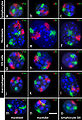
Mouse chromosome territories in different cell types.
-
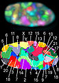
24 chromosome territories in human cells.
Structures of the cell / organelles (TH H1.00.01.2-3) Endomembrane system Cell membrane · Nucleus (and Nucleolus) · Endoplasmic reticulum · Golgi apparatus · Parenthesome · Autophagosome
Vesicles (Exosome · Lysosome · Endosome · Phagosome · Vacuole)
Cytoplasmic granules: Melanosome · Microbody (Glyoxysome, Peroxisome) · Weibel-Palade bodyCytoskeleton Endosymbionts Other internal External Structures of the cell nucleus / nuclear protein Envelope (membrane)/
nuclear laminaNucleolus Other Chromatin · Dot (PML body) · Paraspeckle
SMC protein: Cohesin (SMC1A, SMC1B, SMC3) · Condensin (NCAPD2, NCAPD3, NCAPG, NCAPG2, NCAPH, NCAPH2, SMC2, SMC4) · DNA repair (SMC5, SMC6)
Transition nuclear protein: TNP1, TNP2
Nuclear matrix · Nucleoplasm · Nucleoskeleton · Nucleosol
see also transcription factors and intracellular receptorsCategories:- Organelles
- Cell biology


Wikimedia Foundation. 2010.