- Centriole
-
 Schematic of centriole showing microtubule triplets
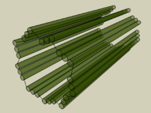 Three-dimensional view of a centriole
 Centrioles from common shore crab hepatopancreas
A Centriole is a barrel-shaped cell structure[1] found in most animal eukaryotic cells, though it is absent in higher plants and most fungi.[2] The walls of each centriole are usually composed of nine triplets of microtubules (protein of the cytoskeleton). Deviations from this structure include Drosophila melanogaster embryos, with nine doublets, and Caenorhabditis elegans sperm cells and early embryos, with nine singlets; [3][4]. Crabs may also exhibit nine doublets, (see picture). An associated pair of centrioles, arranged perpendicularly and surrounded by an amorphous mass of dense material (the pericentriolar material) constitutes the compound structure known as the centrosome.[1] Edouard van Beneden and Theodor Boveri made the first observation and identification of centrioles in 1883 and 1888 respectively, [5][6]while the pattern of centriole replication was first worked out independently by Etienne de Harven and Joseph G. Gall around 1950's [7][8]
Contents
Cell division
Centrioles are involved in the organization of the mitotic spindle and in the completion of cytokinesis.[9] Centrioles were previously thought to be required for the formation of a mitotic spindle in animal cells. However, more recent experiments have demonstrated that cells whose centrioles have been removed via laser ablation can still progress through the G1 stage of interphase before centrioles can be synthesized later in a de novo fashion.[10] Additionally, mutant flies lacking centrioles develop normally, although the adult flies lack flagella and cilia.[11]
Cellular organization
Centrioles are a very important part of centrosomes, which are involved in organizing microtubules in the cytoplasm.[12][13] The position of the centriole determines the position of the nucleus and plays a crucial role in the spatial arrangement of the cell.
Ciliogenesis
In organisms with flagella and cilia, the position of these organelles is determined by the mother centriole, which becomes the basal body. An inability of cells to use centrioles to make functional cilia and flagella has been linked to a number of genetic and developmental diseases. In particular, the inability of centrioles to properly migrate prior to ciliary assembly has recently been linked to Meckel-Gruber syndrome.
Animal development
Proper orientation of cilia via centriole positioning toward the posterior of embryonic node cells is critical for establishing left–right asymmetry during mammalian development.[citation needed]
Centriole duplication
Cells in G0 and G1 usually contain two complete centrioles. The older of the two centrioles in a pair is termed the mother centriole, whereas the younger is termed the daughter centriole. During the cell division cycle, a new centriole grows from the side of each of the existing "mother" centrioles. After centriole duplication, the two pairs of centrioles remain attached to each other in an orthogonal configuration until mitosis, when the mother and daughter centrioles separate in a manner dependent upon the enzyme separase.[14]
The two centrioles in the centrosome are connected to each other by unidentified proteins. The mother centriole has radiating appendages at the distal end of its long axis and is attached to the daughter centriole at the other proximal end. Each daughter cell formed after cell division will inherit one of these pairs (one older and one newer centriole). Duplication of centrioles starts at the time of the G1/S transition and ends before the onset of mitosis.[9]
Origin
The last common ancestor of all eukaryotes was a ciliated cell with centrioles. Some lineages of eukaryotes do not have centrioles anymore, for example land plants. It is unclear if the last common ancestor had one[15] or two cilia.[16] Important genes required for centriole duplication, like centrins, are only found in eukaryotes and neither in eubacteria or archea.[15]
References
- ^ a b Eddé, B; Rossier, J; Le Caer, JP; Desbruyères, E; Gros, F; Denoulet, P (1990). "Posttranslational glutamylation of alpha-tubulin". Science 247 (4938): 83–5. Bibcode 1990Sci...247...83E. doi:10.1126/science.1967194. PMID 1967194.
- ^ Quarmby, LM; Parker, JD (2005). "Cilia and the cell cycle?". The Journal of cell biology 169 (5): 707–10. doi:10.1083/jcb.200503053. PMC 2171619. PMID 15928206. http://www.pubmedcentral.nih.gov/articlerender.fcgi?tool=pmcentrez&artid=2171619.
- ^ Delattre, M; Gönczy, P (2004). "The arithmetic of centrosome biogenesis". Journal of cell science 117 (Pt 9): 1619–30. doi:10.1242/jcs.01128. PMID 15075224.
- ^ Leidel, S; Delattre, M; Cerutti, L; Baumer, K; Gönczy, P (2005). "SAS-6 defines a protein family required for centrosome duplication in C. Elegans and in human cells". Nature cell biology 7 (2): 115–25. doi:10.1038/ncb1220. PMID 15665853.
- ^ Wunderlich, V. (2002). "JMM - Past and Present". Journal of Molecular Medicine 80 (9): 545–548. doi:10.1007/s00109-002-0374-y. PMID 12226736.
- ^ Boveri, Theodor (1888). Zellen-Studien II: Die Befruchtung und Teilung des Eies von Ascaris megalocephala.. Jena: Gustav Fischer Verlag. http://www.biodiversitylibrary.org/item/29952.
- ^ Wolfe, Stephen L. (1977 - First Edition). Biology: the foundations. Wadsworth Pub. Co.. http://books.google.com/books/about/Biology.html?id=Fq8TAQAAIAAJ.
- ^ Vorobjev, I. A.; Nadezhdina, E. S. (1987). "The Centrosome and Its Role in the Organization of Microtubules". International Review of Cytology. International Review of Cytology 106: 227–293. doi:10.1016/S0074-7696(08)61714-3. ISBN 9780123645067. PMID 3294718.. See also de Harven's own recollections of this work: de Harven, Etienne (1994). "Early observations of centrioles and mitotic spindle fibers by transmission electron microscopy". Biol Cell 80 (2–3): 107–109. doi:10.1016/0248-4900(94)90028-0. PMID 8087058. http://www.biolcell.org/boc/080/0107/boc0800107.pdf.
- ^ a b Salisbury, JL; Suino, KM; Busby, R; Springett, M (2002). "Centrin-2 is required for centriole duplication in mammalian cells". Current biology : CB 12 (15): 1287–92. doi:10.1016/S0960-9822(02)01019-9. PMID 12176356.
- ^ La Terra, S; English, CN; Hergert, P; McEwen, BF; Sluder, G; Khodjakov, A (2005). "The de novo centriole assembly pathway in HeLa cells: cell cycle progression and centriole assembly/maturation". The Journal of cell biology 168 (5): 713–22. doi:10.1083/jcb.200411126. PMC 2171814. PMID 15738265. http://www.pubmedcentral.nih.gov/articlerender.fcgi?tool=pmcentrez&artid=2171814.
- ^ Basto, R; Lau, J; Vinogradova, T; Gardiol, A; Woods, CG; Khodjakov, A; Raff, JW (2006). "Flies without centrioles". Cell 125 (7): 1375–86. doi:10.1016/j.cell.2006.05.025. PMID 16814722.
- ^ Feldman, JL; Geimer, S; Marshall, WF (2007). "The mother centriole plays an instructive role in defining cell geometry". PLoS biology 5 (6): e149. doi:10.1371/journal.pbio.0050149. PMC 1872036. PMID 17518519. http://www.pubmedcentral.nih.gov/articlerender.fcgi?tool=pmcentrez&artid=1872036.
- ^ Beisson, J; Wright, M (2003). "Basal body/centriole assembly and continuity". Current opinion in cell biology 15 (1): 96–104. doi:10.1016/S0955-0674(02)00017-0. PMID 12517710.
- ^ Tsou, MF; Stearns, T (2006). "Mechanism limiting centrosome duplication to once per cell cycle". Nature 442 (7105): 947–51. Bibcode 2006Natur.442..947T. doi:10.1038/nature04985. PMID 16862117.
- ^ a b Bornens, M.; Azimzadeh, J. (2007). Origin and Evolution of the Centrosome. "Eukaryotic Membranes and Cytoskeleton". Advances in experimental medicine and biology. Advances in Experimental Medicine and Biology 607: 119–129. doi:10.1007/978-0-387-74021-8_10. ISBN 978-0-387-74020-1. PMID 17977464.
- ^ Rogozin, I. B.; Basu, M. K.; Csuros, M.; Koonin, E. V. (2009). "Analysis of Rare Genomic Changes Does Not Support the Unikont-Bikont Phylogeny and Suggests Cyanobacterial Symbiosis as the Point of Primary Radiation of Eukaryotes". Genome Biology and Evolution 1: 99–113. doi:10.1093/gbe/evp011. PMC 2817406. PMID 20333181. http://www.pubmedcentral.nih.gov/articlerender.fcgi?tool=pmcentrez&artid=2817406.
The centrosome and its components Centrioles Pericentriolar material other proteins Structures of the cell / organelles (TH H1.00.01.2-3) Endomembrane system Cytoskeleton Endosymbionts Other internal External Categories:- Organelles


Wikimedia Foundation. 2010.