- Myofibril
-
Myofibril 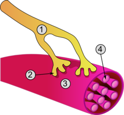
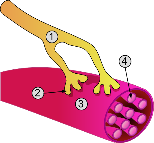
1. Axon
2. Neuromuscular junction
3. Muscle fiber
4. MyofibrilLatin myofibrilla MeSH Myofibrils Code TH H2.00.05.0.00007  A diagram of the structure of a Myofibril
 Sliding filament model of muscle contraction
A myofibril is a basic unit of a muscle. Muscles are composed of tubular cells called myocytes or myofibers. Myofibers are composed of tubular myofibrils. Myofibrils are composed of long proteins such as actin, myosin, and titin, and other proteins that hold them together. These proteins are organized into thin filaments and thick filaments, which repeat along the length of the myofibril in sections called sarcomeres. Muscles contract by sliding the thin (actin) and thick (myosin) filaments along each other.
Actomyosin motors are important in muscle contraction (relying in this case on "classical myosins") as well as other processes like retraction of membrane blebs, filiopod retraction, and uropodium advancement (relying in this case on "nonclassical myosins").
Contents
Structure
The filaments of myofibrils, myofilaments, consist of two types, thick and thin.
- Thin filaments consist primarily of the protein actin, coiled with nebulin filaments.
- Thick filaments consist primarily of the protein myosin, held in place by titin filaments.
The protein complex composed of actin and myosin is sometimes referred to as "actomyosin."
In striated muscle, such as skeletal and cardiac muscle, the actin and myosin filaments each have a specific and constant length on the order of a few micrometers, far less than the length of the elongated muscle cell (a few millimeters in the case of human skeletal muscle cells). The filaments are organized into repeated subunits along the length of the myofibril. These subunits are called sarcomeres. The muscle cell is nearly filled with myofibrils running parallel to each other on the long axis of the cell. The sarcomeric subunits of one myofibril are in nearly perfect alignment with those of the myofibrils next to it. This alignment gives rise to certain optical properties which cause the cell to appear striped or striated. In smooth muscle cells, this alignment is absent, hence there are no apparent striations and the cells are called smooth.
Appearance
The names of the various sub-regions of the sarcomere are based on their relatively lighter or darker appearance when viewed through the light microscope. Each sarcomere is delimited by two very dark colored bands called Z-discs or Z-lines (from the German zwischen meaning between). These Z-discs are dense protein discs that do not easily allow the passage of light. The T-tubule is present in this area. The area between the Z-discs is further divided into two lighter colored bands at either end called the I-bands, and a darker, grayish band in the middle called the A band.
The I bands appear lighter because these regions of the sarcomere mainly contain the thin actin filaments, whose smaller diameter allows the passage of light between them. The A band, on the other hand, contains mostly myosin filaments whose larger diameter restricts the passage of light. A stands for anisotropic and I for isotropic, referring to the optical properties of living muscle as demonstrated with polarized light microscopy.
The parts of the A band that abut the I bands are occupied by the both actin and myosin filaments (where they interdigitate as described above). Also within the A band is a relatively brighter central region called the H-zone (from the German helle, meaning bright) in which there is no actin/myosin overlap when the muscle is in a relaxed state. Finally, the A band is bisected by a dark central line called the M-line (from the German mittel meaning middle).
Action
When a muscle contracts, the actin is pulled along myosin toward the center of the sarcomere until the actin and myosin filaments are completely overlapped. The H zone becomes smaller and smaller due to the increasing overlap of actin and myosin filaments, and the muscle shortens. Thus when the muscle is fully contracted, the H zone is no longer visible (as in the bottom diagram, left). Note that the actin and myosin filaments themselves do not change length, but instead slide past each other. This is known as the sliding filament theory of muscle contraction.
See also
References
External links
- http://www.nismat.org/physcor/muscle.html
- http://msjensen.cehd.umn.edu/1135/Links/Animations/Flash/0008-swf_sarcomere_shor.swf
(animation of sarcomeres contraction)
Structures of the cell / organelles (TH H1.00.01.2-3) Endomembrane system Cytoskeleton Endosymbionts Other internal External B strc: edmb (perx), skel (ctrs), epit, cili, mito, nucl (chro) Histology: muscle tissue (TH H2.00.05, H3.3) Smooth
muscleStriated
muscleCostamere/
DAPCMembrane/
extracellularIntracellularDystrophin · Dystrobrevin (A, B) · Syntrophin (A, B1, B2, G1, G2) · Syncoilin · Dysbindin · Synemin/desmuslin
related: NOS1 · Caveolin 3GeneralNeuromuscular junction · Motor unit · Muscle spindle · Excitation-contraction coupling · Sliding filament mechanismBothFiberCellsOtherOther/
ungroupedCategories:- Organelles


Wikimedia Foundation. 2010.