- MUC1
-
Mucin 1, cell surface associated 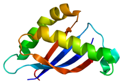
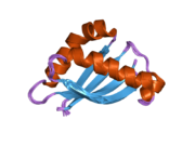
SEA Domain of MUC1. PDB rendering based on 2acm.Available structures PDB 2acm Identifiers Symbols MUC1; CD227; EMA; H23AG; KL-6; MAM6; MUC-1; MUC-1/SEC; MUC-1/X; MUC1/ZD; PEM; PEMT; PUM External IDs OMIM: 158340 MGI: 97231 HomoloGene: 8416 GeneCards: MUC1 Gene Gene Ontology Molecular function • protein binding Cellular component • extracellular region
• nucleus
• cytoplasm
• plasma membrane
• integral to plasma membrane
• cell surface
• apical plasma membraneSources: Amigo / QuickGO RNA expression pattern 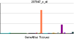
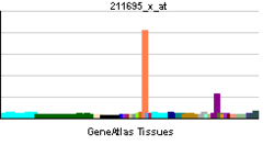
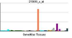
More reference expression data Orthologs Species Human Mouse Entrez 4582 17829 Ensembl ENSG00000185499 ENSMUSG00000042784 UniProt P15941 Q99K60 RefSeq (mRNA) NM_001018016.2 NM_013605.1 RefSeq (protein) NP_001018016.1 NP_038633.1 Location (UCSC) Chr 1:
155.16 – 155.16 MbChr 3:
89.03 – 89.04 MbPubMed search [1] [2] Mucin 1, cell surface associated (MUC1) or polymorphic epithelial mucin (PEM) is a mucin encoded by the MUC1 gene in humans.[1] MUC1 is a proteoglycan with extensive O-linked glycosylation of its extracellular domain. Mucins line the apical surface of epithelial cells in the lungs, stomach, intestines, eyes and several other organs.[2] Mucins protect the body from infection by pathogen binding to oligosaccharides in the extracellular domain, preventing the pathogen from reaching the cell surface.[3] Overexpression of MUC1 is often associated with colon, breast, ovarian, lung and pancreatic cancers.[4]
Contents
Structure
MUC1 is a member of the mucin family and encodes a membrane bound, glycosylated phosphoprotein. MUC1 has a core protein mass of 120-225 kDa which increases to 250-500 kDa with glycosylation. It extends 200-500 nm beyond the surface of the cell.[5]
The protein is anchored to the apical surface of many epithelia by a transmembrane domain. Beyond the transmembrane domain is a SEA domain that contains a cleavage site for release of the large extracellular domain. The release of mucins is performed by sheddases.[6] The extracellular domain includes a 20 amino acid variable number tandem repeat (VNTR) domain, with the number of repeats varying from 20 to 120 in different individuals. These repeats are rich in serine, threonine and proline residues which permits heavy o-glycosylation.[5]
Multiple alternatively spliced transcript variants that encode different isoforms of this gene have been reported, but the full-length nature of only some has been determined.[7]
MUC1 is cleaved in the endoplasmic reticulum into two pieces, the cytoplasmic tail including the transmembrane domain and the extracellular domain. These domain tightly associate in a non-covalent fashion.[8] This tight, non-covalent association is not broken by treatment with urea, low pH, high salt or boiling. Treatment with sodium dodecyl sulfate triggers dissociation of the subunits.[9] The cytoplasmic tail of MUC1 is 72 amino acids long and contains several phosporylation sites.[10]
Function
The protein serves a protective function by binding to pathogens[11] and also functions in a cell signaling capacity.[10]
Overexpression, aberrant intracellular localization, and changes in glycosylation of this protein have been associated with carcinomas. eg. The CanAg tumour antigen is a novel glycoform of MUC1.[12]
Interactions
MUC1 has been shown to interact with HER2/neu,[13][14] Plakoglobin,[13] CTNND1,[15] SOS1[14][16] and Grb2.[16]
Role in Cancer
The ability of chemotherapeutic drugs to access the cancer cells is inhibited by the heavy glycosylation in the extracellular domain of MUC1. The glycosylation creates a highly hydrophilic region which prevents hydrophobic chemotherapeutic drugs from passing through. This prevents the drugs from reaching their targets which usually reside within the cell. Similarly, the glycosylation has been shown to bind to growth factors. This allows cancer cells which produce a large amount of MUC1 to concentrate growth factors near their receptors, increasing receptor activity and the growth of cancer cells. MUC1 also prevents the interaction of immune cells with receptors on the cancer cell surface through steric hindrance. This inhibits an anti-tumor immune response.[2]
Preventing Cell Death
MUC1 cytoplasmic tail has been shown to bind to p53. This interaction is increased by genotoxic stress. MUC1 and p53 were found to be associated with the p53 response element of the p21 gene promoter. This results in activation of p21 which results in cell cycle arrest. Association of MUC1 with p53 in cancer results in inhibition of p53-mediated apoptosis and promotion of p53-mediated cell cycle arrest.[17]
Overexpression of MUC1 in fibroblasts increased the phosphorylation of Akt. Phosphorylation of Akt results in phosphorylation of Bcl-2-associated death promoter. This results in dissociation of Bcl-2-associated death promoter with Bcl-2 and Bcl-xL. Activation was shown to be dependent on the upstream activation of PI3K. Additionally, MUC1 was shown to increase expression of Bcl-xL. Overexpression of of MUC1 in cancer. The presence of free Bcl-2 and Bcl-xL prevents the release of cytochrome c from mitochondria, thereby preventing apoptosis.[18]
MUC1 cytoplasmic tail is shuttled to the mitochondria through interaction with hsp90. This interaction is induced through phosphorylation of the MUC1 cytoplasmic tail by Src (gene). Src is activated by the EGF receptor family ligand Neuregulin. The cytoplasmic tail is then inserted into the mitochondrial outer membrane. Localization of MUC1 to the mitochondria prevents the activation of apoptotic mechanisms.[19]
Promoting Tumor Invasion
MUC1 cytoplasmic tail was shown to interact with Beta-catenin. A SXXXXXSSL motif was identified in MUC1 that is conserved with other beta-catenin binding partners. This interaction was shown to be dependent on cell adhesion.[20] Studies have demonstrated that MUC1 is phosphorylated on a YEKV motif. Phosphorylation of this site has been demonstrated by LYN through mediation of interleukin 7,[21] Src through mediation of EGFR,[22][23] and PRKCD.[24] This interaction is antagonized by degradation of beta-catenin by GSK3B. MUC1 blocks the phosphorylation-dependent degradation of beta-catenin by GSK3B.[25][26] The end result is that increased expression of MUC1 in cancer increases stabilized beta-catenin. This promotes the expression of vimentin and CDH2. These proteins are associated with a mesenchymal phenotype, characterized by increased motility and invasiveness. In cancer cells, increased expression of MUC1 promotes cancer cell invasion through beta-catenin, resulting in the initiation of epithelial-mesenchymal transition which promotes the formation of metastases.[27][28]
See also
References
- ^ Gendler SJ, Lancaster CA, Taylor-Papadimitriou J, Duhig T, Peat N, Burchell J, Pemberton L, Lalani EN, Wilson D (September 1990). "Molecular cloning and expression of human tumor-associated polymorphic epithelial mucin". J. Biol. Chem. 265 (25): 15286–93. PMID 1697589.
- ^ a b Hollingsworth MA, Swanson BJ (January 2004). "Mucins in cancer: protection and control of the cell surface". Nat. Rev. Cancer 4 (1): 45–60. doi:10.1038/nrc1251. PMID 14681689.
- ^ Moncada DM, Kammanadiminiti SJ, Chadee K (July 2003). "Mucin and Toll-like receptors in host defense against intestinal parasites". Trends Parasitol 19 (7): 305–311. doi:10.1016/S1471-4922(03)00122-3. PMID 12855381.
- ^ Gendler SJ (July 2001). "MUC1, the renaissance molecule". J. Mammary Gland Biol Neoplasia 6 (3): 339–353. doi:10.1023/A:1011379725811. PMID 11547902.
- ^ a b Brayman M, Thathiah A, Carson DD (January 2004). "MUC1: a multifunctional cell surface component of reproductive tissue epithelia". Reprod Biol Endocrinol 2: 4. doi:10.1186/1477-7827-2-4. PMC 320498. PMID 14711375. http://www.pubmedcentral.nih.gov/articlerender.fcgi?tool=pmcentrez&artid=320498.
- ^ Hattrup CL, Gendler, SJ (2008). "Structure and Function of the Cell Surface (Tethered) Mucins". Annu. Rev. Physiol. 70: 431–457. doi:10.1146/annurev.physiol.70.113006.100659. PMID 17850209.
- ^ "Entrez Gene: SEA Domain of MUC1". http://www.ncbi.nlm.nih.gov/sites/entrez?Db=gene&Cmd=ShowDetailView&TermToSearch=4582.
- ^ Ligtenberg MJ, Kruijshaar :, Buijs F, van Meijer M, Litvinov SV, Hilkens J (March 1992). "Cell-associated episialin is a complex containing two proteins derived from a common precursor". J. Biol. Chem. 267 (9): 6171–6177. PMID 1556125.
- ^ Julian J, Carson DD (May 2002). "Formation of MUC1 metabolic complex is conserved in tumor-derived and normal epithelial cells". Biochem Biophys Res Commun 293 (4): 1183–1190. doi:10.1016/S0006-291X(02)00352-2. PMID 12054500.
- ^ a b Singh PK, Hollingsworth MA (August 2006). "Cell surface-associated mucins in signal transduction". Trends Cell Biol 16 (9): 467–476. doi:10.1016/j.tcb.2006.07.006. PMID 16904320.
- ^ Linden, SK; Sheng YH, Every AL, Miles KM, Skoog EC, Florin TH, Sutton P, McGuckin MA (Oct. 2009). Van Nhieu, Guy Tran. ed. "MUC1 limits Helicobacter pylori infection both by steric hindrance and by acting as a releasable decoy". PLoS Pathog. 5 (10): e1000617. doi:10.1371/journal.ppat.1000617. PMC 2752161. PMID 19816567. http://www.pubmedcentral.nih.gov/articlerender.fcgi?tool=pmcentrez&artid=2752161.
- ^ http://jco.ascopubs.org/content/21/2/211.full
- ^ a b Li, Yongqing; Yu Wei-Hsuan, Ren Jian, Chen Wen, Huang Lei, Kharbanda Surender, Loda Massimo, Kufe Donald (Aug. 2003). "Heregulin targets gamma-catenin to the nucleolus by a mechanism dependent on the DF3/MUC1 oncoprotein". Mol. Cancer Res. (United States) 1 (10): 765–75. ISSN 1541-7786. PMID 12939402.
- ^ a b Schroeder, J A; Thompson M C, Gardner M M, Gendler S J (Apr. 2001). "Transgenic MUC1 interacts with epidermal growth factor receptor and correlates with mitogen-activated protein kinase activation in the mouse mammary gland". J. Biol. Chem. (United States) 276 (16): 13057–64. doi:10.1074/jbc.M011248200. ISSN 0021-9258. PMID 11278868.
- ^ Li, Y; Kufe D (Feb. 2001). "The Human DF3/MUC1 carcinoma-associated antigen signals nuclear localization of the catenin p120(ctn)". Biochem. Biophys. Res. Commun. (United States) 281 (2): 440–3. doi:10.1006/bbrc.2001.4383. ISSN 0006-291X. PMID 11181067.
- ^ a b Pandey, P; Kharbanda S, Kufe D (Sep. 1995). "Association of the DF3/MUC1 breast cancer antigen with Grb2 and the Sos/Ras exchange protein". Cancer Res. (UNITED STATES) 55 (18): 4000–3. ISSN 0008-5472. PMID 7664271.
- ^ Wei X, Xu H, Kufe D (February 2005). "Human MUC1 oncoprotein regulates p53-responsive gene transcription in the genotoxic stress response". Cancel Cell 7 (2): 167–178. doi:10.1016/j.ccr.2005.01.008. PMID 15710329.
- ^ Raina D, Kharbanda S, Kufe D (May 2004). "The MUC1 oncoprotein activates the anti-apoptotic phosphoinositide 3-kinase/Akt and Bcl-xL pathways in rat 3Y1 fibroblasts". J Biol Chem 279 (20): 20607–20612. doi:10.1074/jbc.M310538200. PMID 14999001.
- ^ Ren J, Bharti A, Raina D, Chen W, Ahmad R, Kufe D (January 2006). "MUC1 oncoprotein is targeted to mitochondria by heregulin-induced activation of c-Src and the molecular chaperone HSP90". Oncogene 25 (1): 20–31. doi:10.1038/sj.onc.1209012. PMID 16158055.
- ^ Yamamoto, M; Bharti A, Li Y, Kufe D (May. 1997). "Interaction of the DF3/MUC1 breast carcinoma-associated antigen and beta-catenin in cell adhesion". J. Biol. Chem. (UNITED STATES) 272 (19): 12492–4. doi:10.1074/jbc.272.19.12492. ISSN 0021-9258. PMID 9139698.
- ^ Li, Yongqing; Chen Wen, Ren Jian, Yu Wei-Hsuan, Li Quan, Yoshida Kiyotsugu, Kufe Donald (2003). "DF3/MUC1 signaling in multiple myeloma cells is regulated by interleukin-7". Cancer Biol. Ther. (United States) 2 (2): 187–93. ISSN 1538-4047. PMID 12750561.
- ^ Li, Y; Kuwahara H, Ren J, Wen G, Kufe D (Mar. 2001). "The c-Src tyrosine kinase regulates signaling of the human DF3/MUC1 carcinoma-associated antigen with GSK3 beta and beta-catenin". J. Biol. Chem. (United States) 276 (9): 6061–4. doi:10.1074/jbc.C000754200. ISSN 0021-9258. PMID 11152665.
- ^ Li, Y; Ren J, Yu W, Li Q, Kuwahara H, Yin L, Carraway K L, Kufe D (Sep. 2001). "The epidermal growth factor receptor regulates interaction of the human DF3/MUC1 carcinoma antigen with c-Src and beta-catenin". J. Biol. Chem. (United States) 276 (38): 35239–42. doi:10.1074/jbc.C100359200. ISSN 0021-9258. PMID 11483589.
- ^ Ren, Jian; Li Yongqing, Kufe Donald (May. 2002). "Protein kinase C delta regulates function of the DF3/MUC1 carcinoma antigen in beta-catenin signaling". J. Biol. Chem. (United States) 277 (20): 17616–22. doi:10.1074/jbc.M200436200. ISSN 0021-9258. PMID 11877440.
- ^ Li, Y; Bharti A, Chen D, Gong J, Kufe D (Dec. 1998). "Interaction of glycogen synthase kinase 3beta with the DF3/MUC1 carcinoma-associated antigen and beta-catenin". Mol. Cell. Biol. (UNITED STATES) 18 (12): 7216–24. ISSN 0270-7306. PMC 109303. PMID 9819408. http://www.pubmedcentral.nih.gov/articlerender.fcgi?tool=pmcentrez&artid=109303.
- ^ Huang L, Chen D, Liu D, Yin L, Kharbanda S, Kufe D (November 2005). "MUC1 oncoprotein blocks glycogen synthase kinase 3beta-mediated phosphorylation and degradation of beta-catenin". Cancer Res 65 (22): 10413–10422. doi:10.1158/0008-5472.CAN-05-2474. PMID 16288032.
- ^ Schroeder, Joyce A; Adriance Melissa C, Thompson Melissa C, Camenisch Todd D, Gendler Sandra J (Mar. 2003). "MUC1 alters beta-catenin-dependent tumor formation and promotes cellular invasion". Oncogene (England) 22 (9): 1324–32. doi:10.1038/sj.onc.1206291. ISSN 0950-9232. PMID 12618757.
- ^ Roy LD, Sahraei M, Subramani DB, Besmer D, Nath S, Tinder TL, Bajaj E, Shanmugam K, Lee YY, Hwang SIL, Gendler SJ, Mukherjee P (March 2011). "MUC1 enhances invasiveness of pancreatic cancer cells by inducing epithelial to mesenchymal transition". Oncogene 30 (12): 1449–1459. doi:10.1038/onc.2010.526. PMC 3063863. PMID 21102519. http://www.pubmedcentral.nih.gov/articlerender.fcgi?tool=pmcentrez&artid=3063863.
Further reading
- Peterson JA, Scallan CD, Ceriani RL, Hamosh M (2002). "Structural and functional aspects of three major glycoproteins of the human milk fat globule membrane". Adv. Exp. Med. Biol. 501: 179–87. PMID 11787681.
- Hu XF, Yang E, Li J, Xing PX (2006). "MUC1 cytoplasmic tail: a potential therapeutic target for ovarian carcinoma". Expert Rev Anticancer Ther 6 (8): 1261–71. doi:10.1586/14737140.6.8.1261. PMID 16925492.
- Leroy X, Buisine MP, Leteurtre E, et al. (2007). "[MUC1 (EMA): A key molecule of carcinogenesis?]". Annales de pathologie 26 (4): 257–66. doi:10.1016/S0242-6498(06)70718-0. PMID 17128152.
- Li Y, Cozzi PJ (2007). "MUC1 is a promising therapeutic target for prostate cancer therapy". Current cancer drug targets 7 (3): 259–71. doi:10.2174/156800907780618338. PMID 17504123.
- Mahanta S, Fessler SP, Park J, Bamdad C (2008). Cordes, Nils. ed. "A Minimal Fragment of MUC1 Mediates Growth of Cancer Cells". PLos ONE 3 (4): e2054. doi:10.1371/journal.pone.0002054. PMC 2329594. PMID 18446242. http://www.pubmedcentral.nih.gov/articlerender.fcgi?tool=pmcentrez&artid=2329594.
- Hikita ST, Kosik KS, Clegg DO, Bamdad C (2008). Cordes, Nils. ed. "MUC1* mediates the growth of human pluripotent stem cells". PLoS ONE 3 (10): e3312. doi:10.1371/journal.pone.0003312. PMC 2553196. PMID 18833326. http://www.pubmedcentral.nih.gov/articlerender.fcgi?tool=pmcentrez&artid=2553196.
- Fessler SP, Wotkowicz MT, Mahanta S, Bamdad C (2009). "MUC1* is a determinant of trastuzumab (Herceptin) resistance in breast cancer cells". Breast Cancer Res Treat 118 (1): 113–24. doi:10.1007/s10549-009-0412-3. PMID 19415485.
PDB gallery External links
This article incorporates text from the United States National Library of Medicine, which is in the public domain.
Mucoproteins OtherProteoglycan Testican · PerlecanChondroitin sulfate proteoglycans: Aggrecan · Neurocan · Brevican · CD44 · CSPG4 · CSPG5 · Platelet factor 4 · Structural maintenance of chromosomes 3Fibromodulin · Lumican · KeratocanOther Activin and inhibin · ADAM · Alpha 1-antichymotrypsin · Apolipoprotein H · CD70 · Asialoglycoprotein · Avidin · B-cell activating factor · 4-1BB ligand · Cholesterylester transfer protein · Clusterin · Colony-stimulating factor · Hemopexin · Lactoferrin · Membrane glycoproteins · Myelin protein zero · Osteonectin · Protein C · Protein S · Serum amyloid P component · Sialoglycoprotein (CD43, Glycophorin, Glycophorin C) · Thrombopoietin · Thyroglobulin · Thyroxine-binding proteins · Transcortin · Tumor necrosis factor-alpha · Uteroglobin · Vitronectinbiochemical families: prot · nucl · carb (glpr, alco, glys) · lipd (fata/i, phld, strd, gllp, eico) · amac/i · ncbs/i · ttpy/i
This membrane protein-related article is a stub. You can help Wikipedia by expanding it.

