- Epithelium
-
This article is about the epithelium as it relates to animal anatomy. For the fungal structure of the same name, see Pileipellis.
Epithelium Code TH H2.00.02.0.00001 Epithelium is one of the four basic types of animal tissue, along with connective tissue, muscle tissue and nervous tissue. Epithelial tissues line the cavities and surfaces of structures throughout the body, and also form many glands. Functions of epithelial cells include secretion, selective absorption, protection, transcellular transport and detection of sensation. In Greek "Epi" means, "on, upon," and "Theli" meaning "tissue." Epithelial layers are avascular, so they must receive nourishment via diffusion of substances from the underlying connective tissue, through the basement membrane.[1][unreliable source?] Epithelia can also be organized into clusters of cells that function as exocrine and endocrine glands. Exocrine and endocrine epithelial cells are highly vascular.
Contents
General structure
Cells in epithelium are very densely packed together like bricks in a wall, leaving very little intercellular space. The cells form continuous sheets which are attached to each other at many locations by tight junctions and desmosomes.[2] The epithelial tissues cover the interior and exterior part of our skin.
Basement membrane
All epithelial cells rest on a basement membrane, which acts as a scaffolding on which epithelium can grow and regenerate after injuries.[3] Epithelial tissue is innervated, but avascular. Thus epithelial tissue must be nourished by substances diffusing from the blood vessels in the underlying tissue. The basement membrane acts as a selectively permeable membrane that determines which substances will be able to enter the epithelium.[4][5]
Cell junctions
Cell junctions are especially abundant in epithelial tissues. They consist of protein complexes and provide contact between neighbouring cells, between a cell and the extracellular matrix, or they build up the paracellular barrier of epithelia and control the paracellular transport.[citation needed]
Cell junctions are the contact points between plasma membrane and tissue cells. There are mainly 5 different types of cell junctions. They are tight junctions, adherens junctions, desmosomes, hemidesmosomes, and gap junctions. Tight junctions are a pair of trans-membranar protein fused on outer plasma membrane. Adherens junctions are a plaque (protein layer on the inside plasma membrane) which attaches both protein and microfilaments. Desmosomes attach to the microfilaments of cytoskeleton made up of keratin protein. Hemidesmosomes resemble desmsomes on a section. They are made up of the integrin (a transmembraner protein) instead of cadherin. They attach the epithelial cell to the basement membrane. Gap junctions connect the cytoplasm of two cells and are made up of proteins called connexins (six of which come together to make a connexon).
Classification of epithelial tissue
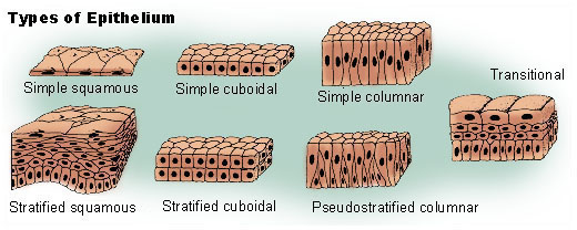
 Types of epithelium
Tissues are generally classified by the morphology of their cells, and the number of layers they are composed of.[2][4][6] Epithelial tissue that is only one cell thick is known as simple epithelium.[7] If it is two or more cells thick, it is known as stratified epithelium.[8] However, when taller simple epithelial cells (see columnar, below) are viewed in cross section with several nuclei appearing at different heights, they can be confused with stratified epithelia. This kind of epithelium is therefore described as "pseudostratified" epithelium.[9]
Simple epithelium
Simple epithelium is one cell thick, that is, every cell is in direct contact with the underlying basement membrane. It is generally found where absorption and filtration occur. The thinness of the epithelial barrier facilitates these processes.[2]
Simple epithelial tissues are generally classified by the shape of their cells. The four major classes of simple epithelium are: (1) simple squamous; (2) simple cuboidal; (3) simple columnar; (4) pseudostratified.[2]
(1) simple squamous; which is found lining areas where passive diffusion of gases occur. e.g walls of capillaries, linings of the pericardial, pleural,and peritoneal cavities, as well as the linings of the alveoli of the lungs.
(2) simple cuboidal: these cells may have secretory, absorptive, or excretory functions. examples include small collecting ducts of kidney,pancreas and salivary gland.
(3) simple columnar; found in areas with extremely high secretive (as in wall of the stomach), or absorptive (as in small intestine) areas. they possess cellular extensions (e.g microvilli in the small intestine, or cilia found almost exclusively in the female reproductive tract).
(4) pseudostratified epithelia; they are also called respiratory epithelium. this is due to their almost exclusive confinement to the larger respiratory airways i.e the nasal cavity, trachea, bronchi e.t.c.
Type Description Squamous Squamous cells have the appearance of thin, flat plates. They fit closely together in tissues; providing a smooth, low-friction surface over which fluids can move easily. The shape of the nucleus usually corresponds to the cell form and helps to identify the type of epithelium. Squamous cells tend to have horizontally flattened, elliptical (oval or shaped like an egg) nuclei because of the thin flattened form of the cell. Classically, squamous epithelia are found lining surfaces utilizing simple passive diffusion such as the alveolar epithelium in the lungs. Specialized squamous epithelia also form the lining of cavities such as the blood vessels (endothelium) and pericardium (mesothelium) and the major cavities found within the body. Cuboidal As their name implies, cuboidal cells are roughly cuboidal in shape, appearing square in cross section. Each cell has a spherical nucleus in the centre. Cuboidal epithelium is commonly found in secretive or absorptive tissue: for example the (secretive) exocrine gland the pancreas and the (absorptive) lining of the kidney tubules as well as in the ducts of the glands. They also constitute the germinal epithelium that covers the female ovary. Columnar Columnar epithelial cells are elongated and column-shaped. Their nuclei are elongated and are usually located near the base of the cells. Columnar epithelium forms the lining of the stomach and intestines. Some columnar cells are specialized for sensory reception such as in the nose, ears and the taste buds of the tongue. Goblet cells (unicellular glands) are found between the columnar epithelial cells of the duodenum. They secrete mucus, which acts as a lubricant. Pseudostratified These are simple columnar epithelial cells whose nuclei appear at different heights, giving the misleading (hence "pseudo") impression that the epithelium is stratified when the cells are viewed in cross section. Pseudostratified epithelium can also possess fine hair-like extensions of their apical (luminal) membrane called cilia. In this case, the epithelium is described as "ciliated" pseudostratified epithelium. Cilia are capable of energy dependent pulsatile beating in a certain direction through interaction of cytoskeletal microtubules and connecting structural proteins and enzymes. The wafting effect produced causes mucus secreted locally by the goblet cells (to lubricate and to trap pathogens and particles) to flow in that direction (typically out of the body). Ciliated epithelium is found in the airways (nose, bronchi), but is also found in the uterus and Fallopian tubes of females, where the cilia propel the ovum to the uterus. Stratified epithelium
Stratified epithelium differs from simple epithelium in that it is multilayered. It is therefore found where body linings have to withstand mechanical or chemical insult such that layers can be abraded and lost without exposing subepithelial layers. Cells flatten as the layers become more apical, though in their most basal layers the cells can be squamous, cuboidal or columnar.[citation needed]
Stratified epithelial tissue also differs from simple epithelial tissue in that stratified epithelial tissues do not contain junctional complexes, and have their cells bound together only by desmosomes.[8]
Stratified epithelia (of columnar, cuboidal or squamous type) can have the following specializations:[citation needed]
Specialization Description Keratinized In this particular case, the most apical layers (exterior) of cells are dead and lose their nucleus and cytoplasm, instead contain a tough, resistant protein called keratin. This specialization makes the epithelium waterproof, so is found in the mammalian skin. The lining of the esophagus is an example of a non-keratinized or "moist" stratified epithelium.[citation needed] Transitional Transitional epithelia are found in tissues that stretch and it can appear to be stratified cuboidal when the tissue is not stretched or stratified squamous when the organ is distended and the tissue stretches. It is sometimes called the urothelium since it is almost exclusively found in the bladder, ureters and urethra.[citation needed] Functions
The primary functions of epithelial tissues are: (1) to protect the tissues that lie beneath it from radiation, desiccation, toxins, and physical trauma; (2) the regulation and exchange of chemicals between the underlying tissues and a body cavity; (3) the secretion of hormones into the blood vascular system, and/or (3) the secretion of sweat, mucus, enzymes, and other products that are delivered by ducts glandular epithelium.[10]
Secretory epithelia
As stated above, secretion is one major function of epithelial cells. Glands are formed from the invagination / infolding of epithelial cells and subsequent growth in the underlying connective tissue. There are two major classifications of glands: endocrine glands and exocrine glands. Endocrine glands secrete their product into the extracellular space where it is rapidly taken up by the blood vascular system. the exocrine glands secrete their products into a duct that then delivers the product to the lumen of an organ or onto the free surface of the epithelium.
Sensing the extracellular environment
"Some epithelial cells are ciliated, and they commonly exist as a sheet of polarised cells forming a tube or tubule with cilia projecting into the lumen." Primary cilia on epithelial cells provide chemosensation, thermosensation and mechanosensation of the extracellular environment by playing "a sensory role mediating specific signalling cues, including soluble factors in the external cell environment, a secretory role in which a soluble protein is released to have an effect downstream of the fluid flow, and mediation of fluid flow if the cilia are motile."[11]
Embryological development
In general, there are epithelial tissues deriving from all of the embryological germ layers[citation needed]:
- from ectoderm (e.g., the epidermis);
- from endoderm (e.g., the lining of the gastrointestinal tract);
- from mesoderm (e.g., the inner linings of body cavities).
However, it is important to note that pathologists do not consider endothelium and mesothelium (both derived from mesoderm) to be true epithelium. This is because such tissues present very different pathology. For that reason, pathologists label cancers in endothelium and mesothelium sarcomas, whereas true epithelial cancers are called carcinomas. Also, the filaments that support these mesoderm-derived tissues are very distinct. Outside of the field of pathology, it is, in general, accepted that the epithelium arises from all three germ layers.[citation needed]
Growing in culture
When growing epithelium in culture, one can determine whether or not a particular cell is epithelial by examining its morphological characteristics. Epithelial cells tend to cluster together, and have a "characteristic tight pavementlike appearance". But this is not always the case, such as when the cells are derived from a tumor. In these cases, it is often necessary to use certain biochemical markers to make a positive identification. The intermediate filament proteins in the cytokeratin group are almost exclusively found in epithelial cells, and so are often used for this purpose.[12]
Location
Epithelium lines both the outside (skin) and the inside cavities and lumen of bodies. The outermost layer of our skin is composed of dead stratified squamous, keratinized epithelial cells.[citation needed]
Tissues that line the inside of the mouth, the esophagus and part of the rectum are composed of nonkeratinized stratified squamous epithelium. Other surfaces that separate body cavities from the outside environment are lined by simple squamous, columnar, or pseudostratified epithelial cells. Other epithelial cells line the insides of the lungs, the gastrointestinal tract, the reproductive and urinary tracts, and make up the exocrine and endocrine glands. The outer surface of the cornea is covered with fast-growing, easily-regenerated epithelial cells. Endothelium (the inner lining of blood vessels, the heart, and lymphatic vessels) is a specialized form of epithelium. Another type, mesothelium, forms the walls of the pericardium, pleurae, and peritoneum.[citation needed]
[editing help required: the internal wiki-link 'gastric epithelium' in the column 'Subtype' of the table below is a circular reference back to this page.]
System Tissue Epithelium Subtype circulatory blood vessels Simple squamous endothelium digestive ducts of submandibular glands Stratified columnar - digestive attached gingiva Stratified squamous, keratinized - digestive dorsum of tongue Stratified squamous, keratinized - digestive hard palate Stratified squamous, keratinized - digestive oesophagus Stratified squamous, non-keratinized - digestive stomach Simple columnar, non-ciliated gastric epithelium digestive small intestine Simple columnar, non-ciliated intestinal epithelium digestive large intestine Simple columnar, non-ciliated intestinal epithelium digestive rectum Simple columnar, non-ciliated - digestive anus Stratified squamous, non-keratinized superior to Hilton's white line
Stratified squamous, keratinized inferior to Hilton's white line- digestive gallbladder Simple columnar, non-ciliated - endocrine thyroid follicles Simple cuboidal - nervous ependyma Simple cuboidal - lymphatic lymph vessel Simple squamous endothelium integumentary skin - dead superficial layer Stratified squamous, keratinized - integumentary sweat gland ducts Stratified cuboidal - integumentary mesothelium of body cavities Simple squamous mesothelium reproductive - female ovaries Simple cuboidal germinal epithelium (female) reproductive - female Fallopian tubes Simple columnar, ciliated - reproductive - female endometrium (uterus) Simple columnar, ciliated - reproductive - female cervix (endocervix) Simple columnar - reproductive - female cervix (ectocervix) Stratified squamous, non-keratinized - reproductive - female vagina Stratified squamous, non-keratinized - reproductive - female labia majora Stratified squamous, keratinized - reproductive - male tubuli recti Simple cuboidal germinal epithelium (male) reproductive - male rete testis Simple cuboidal - reproductive - male ductuli efferentes Pseudostratified columnar - reproductive - male epididymis Pseudostratified columnar, with stereocilia - reproductive - male vas deferens Pseudostratified columnar - reproductive - male ejaculatory duct Simple columnar - reproductive - male (gland) bulbourethral glands Simple columnar - reproductive - male (gland) seminal vesicle Pseudostratified columnar - respiratory oropharynx Stratified squamous, non-keratinized - respiratory larynx Pseudostratified columnar, ciliated respiratory epithelium respiratory larynx - True vocal cords Stratified squamous, non-keratinized - respiratory trachea Pseudostratified columnar, ciliated respiratory epithelium respiratory respiratory bronchioles Simple cuboidal - sensory cornea Stratified squamous, non-keratinized corneal epithelium sensory nose Pseudostratified columnar olfactory epithelium urinary kidney - proximal convoluted tubule Simple cuboidal, with microvilli - urinary kidney - ascending thin limb Simple squamous - urinary kidney - distal convoluted tubule Simple cuboidal, without microvilli - urinary kidney - collecting duct Simple cuboidal - urinary renal pelvis Transitional urothelium urinary ureter Transitional urothelium urinary urinary bladder Transitional urothelium urinary prostatic urethra Transitional urothelium urinary membranous urethra Pseudostratified columnar, non-ciliated - urinary penile urethra Pseudostratified columnar, non-ciliated - urinary external urethral orifice Stratified squamous - Additional images
-
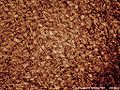
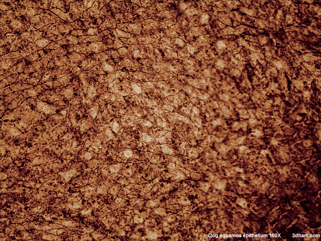
Squamous Epithelium 100x
-
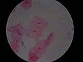
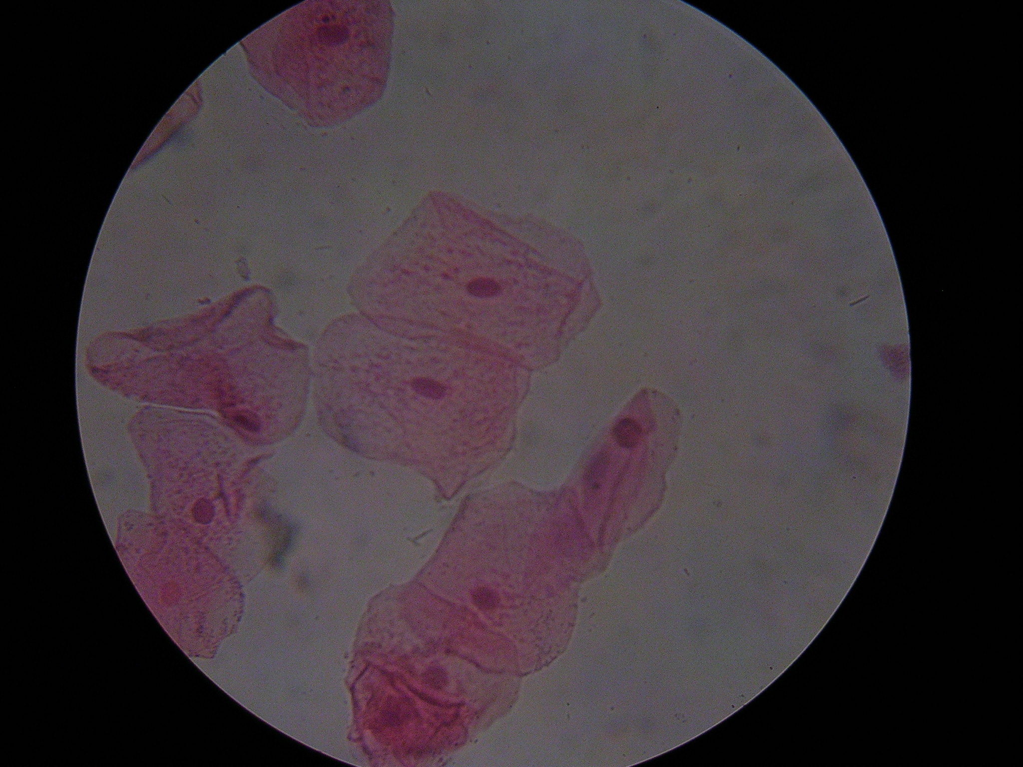
Human cheek cells (Nonkeratinized stratified squamous epithelium) 500x
See also
References
Notes
- ^ "Blue Histology". http://www.lab.anhb.uwa.edu.au/mb140/. Retrieved 2008-12-12.[unreliable source?]
- ^ a b c d Marieb, Elaine M. (1995). Human Anatomy and Physiology (3rd ed.). Benjamin/Cummings. pp. 103–104. ISBN 0-8053-4281-8.
- ^ csxcasdfrg4y24q5qdwsedrMcConnell, Thomas H. (2006). The nature of disease: pathology for the health professions. Lippincott Williams & Wilkins. p. 55. ISBN 9780781753173. http://books.google.com/books?id=chs_lilPFLwC&pg=PA55.
- ^ a b Eurell, Jo Ann C. et al., ed (2006). Dellmann's textbook of veterinary histology. Wiley-Blackwell. p. 18. ISBN 9780781741484. http://books.google.com/books?id=FnS4uiOlRT0C&pyg=PA18.
- ^ Freshney, 2002: p. 3
- ^ Platzer, Werner (2008). Color atlas of human anatomy: Locomotor system. Thieme. p. 8. ISBN 9783135333069. http://books.google.com/books?id=T9bb4T422j8C&pg=PA8.
- ^ van Lommel, 2002: p. 94
- ^ a b van Lommel, 2002: p. 97
- ^ Melfi, Rudy C. & Alley, Keith E., ed (2000). Permar's oral embryology and microscopic anatomy: a textbook for students in dental hygiene. Lippincott Williams & Wilkins. p. 9. ISBN 9780683306446. http://books.google.com/books?id=fGonan0UwhQC&pg=PA9.
- ^ van Lommel, 2002: p. 91
- ^ Adams, M.; Smith, U.M.; Logan, C.V.; Johnson, C.A. (2008). "Recent advances in the molecular pathology, cell biology and genetics of ciliopathies". Journal of Medical Genetics 45 (5): 257–267. doi:10.1136/jmg. PMID 18178628. http://jmg.bmj.com/cgi/content/full/45/5/257.
- ^ Freshney, 2002: p. 9
Bibliography
- Freshney, R.I. (2002). "Introduction". In Freshney, R. Ian & Freshney, Mary. Culture of epithelial cells. John Wiley & Sons. ISBN 9780471401216. http://books.google.com/books?id=KqKNxeWlU6MC&pg=PA1.
- van Lommel, Alfons T.L. (2002). From cells to organs: a histology textbook and atlas. Springer. ISBN 9781402072574. http://books.google.com/books?id=EvYjLNKLu9sC.
Further reading
- Green H (September 2008). "The birth of therapy with cultured cells". Bioessays 30 (9): 897–903. doi:10.1002/bies.20797. PMID 18693268.
- Kefalides, Nicholas A. & Borel, Jacques P., ed (2005). Basement membranes: cell and molecular biology. Gulf Professional Publishing. ISBN 9780121533564. http://books.google.com/books?id=RM-FVY47NEgC.
- Nagpal R, Patel A, Gibson MC (March 2008). "Epithelial topology". Bioessays 30 (3): 260–6. doi:10.1002/bies.20722. PMID 18293365.
- Yamaguchi Y, Brenner M, Hearing VJ (September 2007). "The regulation of skin pigmentation" (Review). J. Biol. Chem. 282 (38): 27557–61. doi:10.1074/jbc.R700026200. PMID 17635904. http://www.jbc.org/cgi/pmidlookup?view=long&pmid=17635904.
Animals Plants Histology: Epithelial proteins (TH H1.00.01.1) Lateral/cell-cell Cell adhesion molecules: Adherens junction (Cadherin) · Desmosome (Desmoglein)
Ion channels: Gap junction/Connexon (Connexin)
Cytoskeleton: Desmosome (Desmoplakin, Plakoglobin, Tonofibril)
other membrane proteins: Tight junction (Claudin, Occludin, MARVELD2)Basal/cell-matrix Apical Categories:- Tissues
Wikimedia Foundation. 2010.