- Olfactory epithelium
-
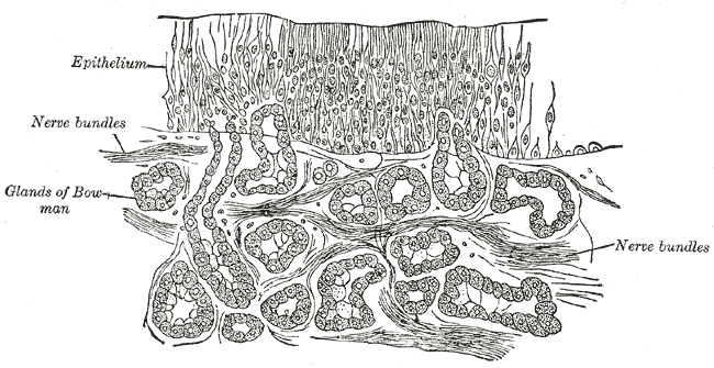
Olfactory epithelium 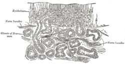
Section of the olfactory mucous membrane. 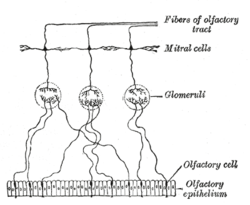
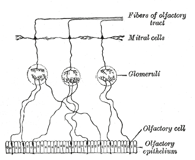
Plan of olfactory neurons. Gray's subject #223 996 MeSH Olfactory+Mucosa Code TH H3.11.07.0.01001 The olfactory epithelium is a specialized epithelial tissue inside the nasal cavity that is involved in smell. In humans, it measures about 1 square centimetre (on each side) and lies on the roof of the nasal cavity about 7 cm above and behind the nostrils.[1] The olfactory epithelium is the part of the olfactory system directly responsible for detecting odors.
Contents
Layers of olfactory epithelium
Olfactory epithelium consists of four distinct cell types: [2]
- Olfactory cells
- Supporting cells
- Basal cells
- Brush Cells
Olfactory cells
The olfactory cells of the epithelium are bipolar neurons which congregate to form the olfactory nerve (cranial nerve I). The apical poles of these neurons are covered with non-motile cilia, with the plasma membrane containing odorant-binding proteins acting as olfactory receptors. The incoming odorants are made soluble by the serous secretion from Bowman's Glands, located in the lamina propria of the mucosa. [3]
Supporting cells
Analogous to neural glial cells, the supporting cells (a.k.a. Sustentacular cells) of the olfactory epithelium function as metabolic and physical support for the olfactory cells. Histologically, the supporting cells are tall columnar cells featuring microvilli and a prominent terminal web. The nuclei of supporting cells are more apically located than those of the other olfactory epithelial cells.
Basal cells
Resting on the basal lamina of the olfactory epithelium, basal cells are stem cells capable of division and differentiation into either supporting or olfactory cells. The constant divisions of the basal cells leads to the olfactory epithelium being replaced every 2–4 weeks.
Basal cells can be divided on the basis of cellular anatomy histological markers into two populations: the horizontal basal cells which line the olfactory epithelium and the slightly more superficial globose basal cells.[4] Horizontal basal cells are now thought to be the primary stem cell population supplying new cells in this system.,[5] although this is subject to some debate with some scientists maintaining that the globose basal cells are the true stem cells.
Brush Cells
A microvilli-bearing columnar cell with basal surface in contact with afferent nerve endings, specialised for transduction of general sensation. Nerve fibres are terminal branches of trigeminal nerve (cranial nerve V), rather than the olfactory nerve, as afferent olfactory signals are.
Bowman's (Olfactory) Glands
Tubuloalveolar serous secreting glands lying in the lamina propria of the mucosa. These glands deliver a proteinaceous secretion via ducts onto the surface of the mucosa. The role of the secretions are to trap and dissolve odiferous substances for the bipolar neurons. Constant flow from the Bowman's glands allows old odors to be constantly washed away. [6]
Pathology
The olfactory epithelium can be damaged by inhalation of toxic fumes, physical injury to the interior of the nose, and possibly by the use of some nasal sprays. Because of its regenerative capacity, damage to the olfactory epithelium can be temporary but in extreme cases, injury can be permanent, leading to anosmia.
Additional images
-

Composition of the Olfactory receptor neuron (captions in German)
-

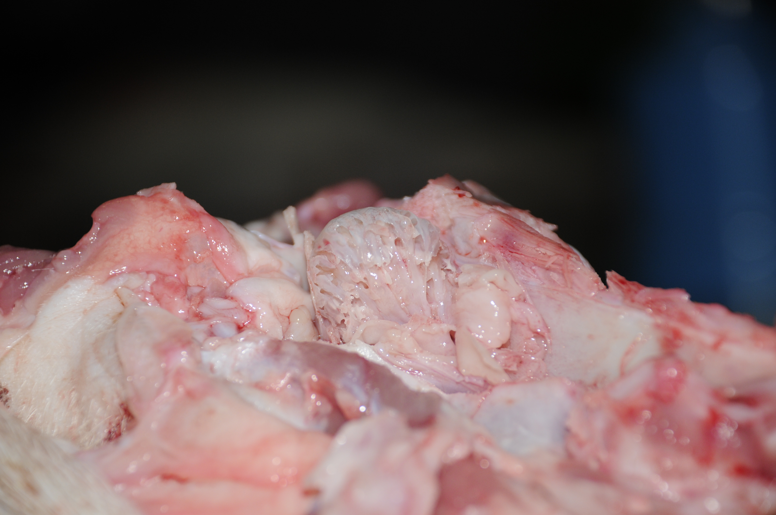
olfactory epithelium pig
References
- ^ Moran, David T.; Rowley Jc, 3rd; Jafek, BW; Lovell, MA (1982), "The fine structure of the olfactory mucosa in man", Journal of Neurocytology 11 (5): 721–746, doi:10.1007/BF01153516, PMID 7143026
- ^ Ross, MH, Histology: A Text and Atlas, 5th Edition. Philadelphia: Lippincott, Williams and Wilkins, 2006. page 615-616.
- ^ Ross, MH, Histology: A Text and Atlas, 5th Edition. Philadelphia: Lippincott, Williams and Wilkins, 2006. page 616.
- ^ Schwob, James E. (2002), "Neural Regeneration and the Peripheral Olfactory System", The Anatomical Record 269 (1): 33–49, doi:10.1002/ar.10047, PMID 11891623
- ^ Leung,C.T.; Coulombe,P.A.; Reed,R.R. (2007) “Contribution of olfactory neural stem cells to tissue maintenance and regeneration”. Nat Neurosci. 10(6):673-4. PMID: 17468753
- ^ Ross, MH, Histology: A Text and Atlas, 5th Edition. Philadelphia: Lippincott, Williams and Wilkins, 2006. page 616.
External links
Sensory system: Olfactory system / Olfaction / Rhinencephalon (TA 15.1, GA 10.992) Olfactory epithelium Olfactory nerve: 1° neuron Olfactory nerve: 2° neuron Lateral olfactory stria/
Primary olfactory cortexPiriform cortex · EC-hippocampus system (Entorhinal cortex, Hippocampal formation) · Prepyriform area · Periamygdaloid cortex
Stria medullaris → Habenular nuclei
Amygdala → Stria terminalis → Hypothalamus
Medial forebrain bundle → HypothalamusMedial olfactory stria Epithelial cells Surface epithelium Simple squamous epithelium (Endothelium, Mesothelium)
Pseudostratified columnar epithelium (Respiratory epithelium)
Stratified squamous epithelium
Stratified cuboidal epithelium
Stratified columnar epithelium
Transitional epithelium/UrotheliumGland/
glandular epitheliumMechanismShapeSecretionSerous glands · Mucous glandsComponentssee also Template:Epithelial neoplasmsCategories:

Wikimedia Foundation. 2010.