- Glomerulus (olfaction)
-
This article is about the structure in the olfactory bulb. For the structure in the kidney, see Glomerulus.
Glomerulus (olfaction) 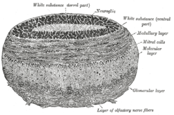
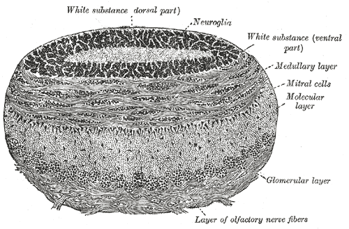
Coronal section of olfactory bulb. 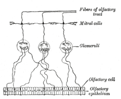
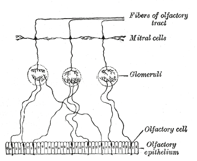
Plan of olfactory neurons. Gray's subject #189 848 The glomerulus (plural glomeruli) is a spherical structure located in the olfactory bulb of the brain where synapses form between the terminals of the olfactory nerve and the dendrites of mitral, periglomerular and tufted cells. Each glomerulus is surrounded by a heterogeneous population of juxtaglomerular neurons (that include periglomerular, short axon, and external tufted cells) and glial cells.[1][2][3] All glomeruli are located near the surface of the olfactory bulb. They are the initial sites for synaptic processing of odor information coming from the nose. A glomerulus is made up of a globular tangle of axons from the olfactory receptor neurons, and dendrites from the mitral and tufted cells, as well as, from cells that surround the glomerulus such as the external tufted cells, periglomerular cells, short axon cells, and astrocytes. In mammals, glomeruli typically range between 50-120 µm in diameter and number between 1800 and 2400 depending on the species.[1][2] Each glomerulus is composed of two compartments, the olfactory nerve zone and the non-olfactory nerve zone. The olfactory nerve zone is composed of preterminals and terminals of the olfactory nerve and is where the olfactory receptor cells make synapses on their targets.[2] The non-olfactory nerve zone is composed of the dendritic processes of intrinsic neurons and is where dendrodendritic interactions between intrinsic neurons occur.[2]
Anatomy
Glomeruli are important waystations in the pathway from the nose to the olfactory cortex and have been found to be critical for odorant signal transduction. The olfactory receptor neurons (ORN), which originate in the nasal epithelium express only one type of olfactory receptor (OR). These ORNs then project their axons to the olfactory bulb. In the olfactory bulb, the ORNs synapse with termination in the glomeruli.[4] Each glomerulus receives input from olfactory receptor neurons expressing only one type of olfactory receptor. The glomerular activation patterns within the olfactory bulb are thought to represent the quality of the odor being detected. These activation patterns of glomeruli can change due to changes in airflow rate and odor concentration in the mucus layer of the nasal cavity.[5] The current dogma is that axons from all ORNs expressing the same receptor converge onto one or two glomeruli of a possible 1800 glomeruli in each olfactory bulb.[4] As the axons of the ORNs migrate towards their specific glomeruli they often overshoot into neighboring glomeruli. Thus, a glomerulus representing a specific OR develops slowly and involves considerable axonal reorganization in order to achieve the highly topographical projection observed in adult mice.[6]
Function
The glomerulus is the basic unit in the odor map of the olfactory bulb. Each odor activates a different pattern of glomeruli, such that, simply by analyzing the different sets of activated glomeruli, one could, in theory, decode the identity of the odor. This odor map, however, is modified by the circuitry within the olfactory bulb so that the spiking pattern of the second-order mitral cells is usually different from that of the olfactory sensory neurons.[7]
References
- ^ a b A. J. Pinching, T. P. S. Powell. (1971) The neuropil of the glomeruli of the olfactory bulb. J. Cell Sci. 9: 347-377. PMID 4108057
- ^ a b c d K. Kosaka, K. Toida, Y. Aika, T. Kosaka. (1998) How simple is the organization of the olfactory glomerulus?: the heterogeneity of so-called periglomerular cells. Neuroscience Research 30: 101-110. PMID 9579643
- ^ "Wachowiak and Shipley (2006) Coding and synaptic processing of sensory information in the glomerular layer of the olfactory bulb. Semin Cell Dev Biol. 17(4):411-23 PMID: 16765614". ScienceDirect. http://www.sciencedirect.com/science?_ob=ArticleURL&_udi=B6WX0-4JWDY4P-6&_user=418620&_coverDate=08/31/2006&_rdoc=1&_fmt=high&_orig=gateway&_origin=gateway&_sort=d&_docanchor=&view=c&_acct=C000019840&_version=1&_urlVersion=0&_userid=418620&md5=c8836deed46bf4d7f5d7f29a2da0714e&searchtype=a#secx2. Retrieved 2011-03-10.
- ^ a b S.J. Royal and B. Key (1999) Development of P2 olfactory glomeruli in P2-internal ribosome entry site-Tau-LacZ transgenic mice. J. Neurosci 19: 9856-9864 PMID 10559395
- ^ Y. Oka, Y. Taki, K. Touhara (2009) Nasal airflow rate affects the sensitivity and pattern of glomerular odorant responses in the mouse olfactory bulb. J. Neurosci 29: 12070-12078 PMID 19793965
- ^ "M.S. Potter, C. Zheng, S.K. David, P. Feinstein, E. F. Scott, and P. Mombaerts (2001) Structure and emergence of specific olfactory glomeruli in the mouse J. Neurosci 21: 9713-9723 PMID 11739580". 2001-12-15. http://www.jneurosci.org/cgi/content/short/21/24/9713. Retrieved 2010-04-27.
- ^ "Friedrich and Laurent (2001) Dynamic Optimization of Odor Representations by Slow Temporal Patterning of Mitral Cell Activity Science 2001 Feb 2;291(5505):889-94. 17(4):411-23 PMID: 11157170". Science. http://www.sciencemag.org/content/291/5505/889.long. Retrieved 2011-03-10.
Sensory system: Olfactory system / Olfaction / Rhinencephalon (TA 15.1, GA 10.992) Olfactory epithelium Olfactory nerve: 1° neuron Olfactory nerve: 2° neuron Lateral olfactory stria/
Primary olfactory cortexPiriform cortex · EC-hippocampus system (Entorhinal cortex, Hippocampal formation) · Prepyriform area · Periamygdaloid cortex
Stria medullaris → Habenular nuclei
Amygdala → Stria terminalis → Hypothalamus
Medial forebrain bundle → HypothalamusMedial olfactory stria Categories:
Wikimedia Foundation. 2010.