- Kidney
-
For other uses, see Kidney (disambiguation).
Kidney 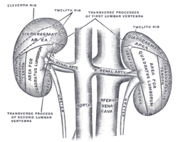
Human kidneys viewed from behind with spine removed Latin ren Artery renal artery Vein renal vein Nerve renal plexus 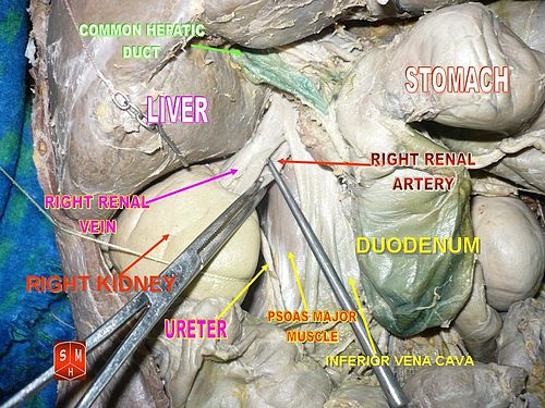 Right kidney
 Left kidney
The kidneys, organs with several functions, serve essential regulatory roles in most animals, including vertebrates and some invertebrates. They are essential in the urinary system and also serve homeostatic functions such as the regulation of electrolytes, maintenance of acid–base balance, and regulation of blood pressure (via maintaining salt and water balance). They serve the body as a natural filter of the blood, and remove wastes which are diverted to the urinary bladder. In producing urine, the kidneys excrete wastes such as urea and ammonium, and they are also responsible for the reabsorption of water, glucose, and amino acids. The kidneys also produce hormones including calcitriol, erythropoietin, and the enzyme renin.
Located at the rear of the abdominal cavity in the retroperitoneum, the kidneys receive blood from the paired renal arteries, and drain into the paired renal veins. Each kidney excretes urine into a ureter, itself a paired structure that empties into the urinary bladder.
Renal physiology is the study of kidney function, while nephrology is the medical specialty concerned with kidney diseases. Diseases of the kidney are diverse, but individuals with kidney disease frequently display characteristic clinical features. Common clinical conditions involving the kidney include the nephritic and nephrotic syndromes, renal cysts, acute kidney injury, chronic kidney disease, urinary tract infection, nephrolithiasis, and urinary tract obstruction.[1] Various cancers of the kidney exist; the most common adult renal cancer is renal cell carcinoma. Cancers, cysts, and some other renal conditions can be managed with removal of the kidney, or nephrectomy. When renal function, measured by glomerular filtration rate, is persistently poor, dialysis and kidney transplantation may be treatment options. Although they are not severely harmful, kidney stones can be a pain and a nuisance. The removal of kidney stones includes sound wave treatment to break up the stones into smaller pieces, which are then passed through the urinary tract. One common symptom of kidney stones is a sharp pain in the medial/lateral segments of the lower back.
Contents
Anatomy
Location
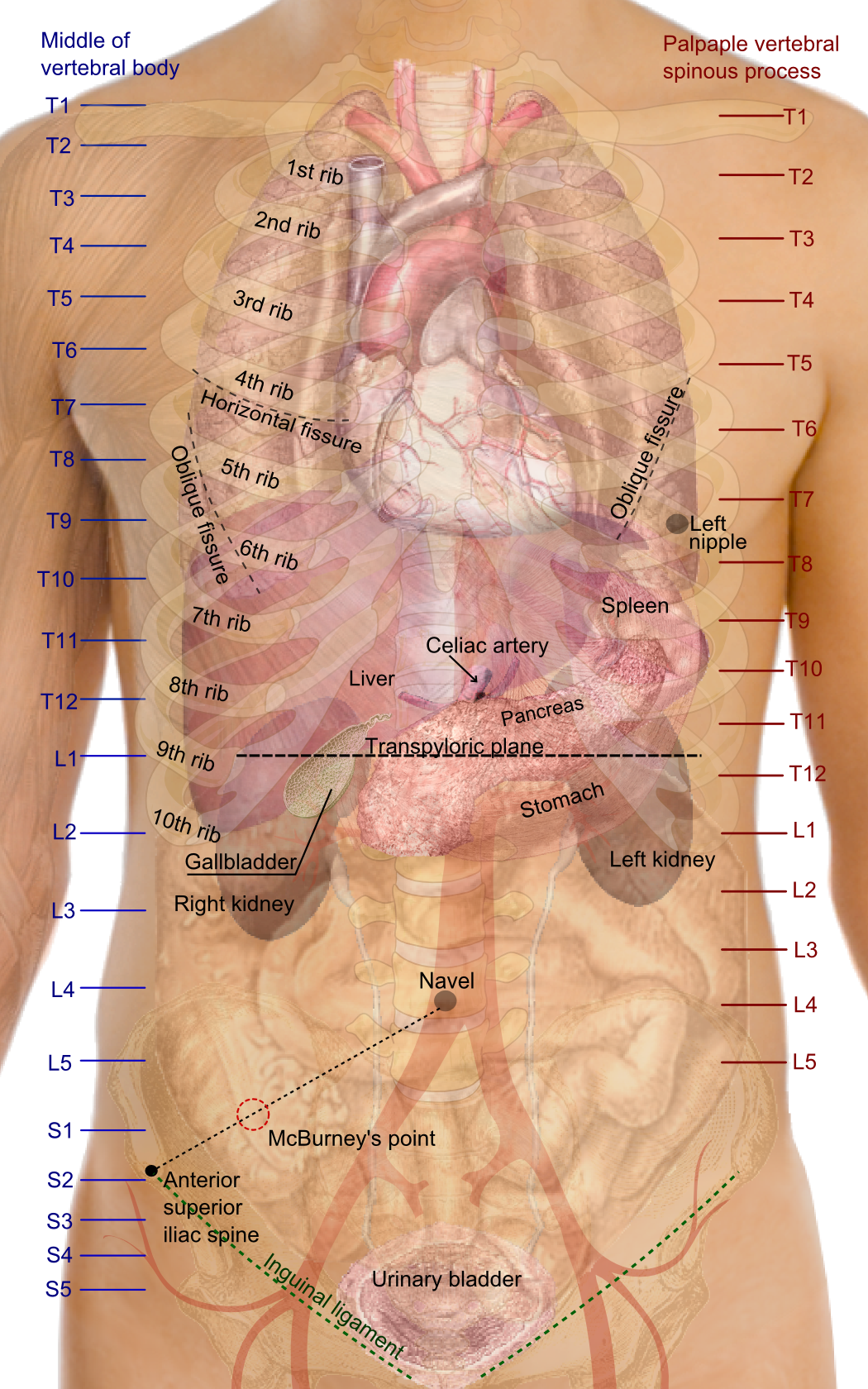
 Surface projections of the organs of the trunk, showing kidneys at the level of T12 to L3.
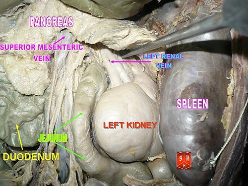
In humans the kidneys are located in the abdominal cavity, more specifically in the paravertebral gutter and lie in a retroperitoneal position at a slightly oblique angle. There are two, one on each side of the spine. The asymmetry within the abdominal cavity caused by the liver typically results in the right kidney being slightly lower than the left, and left kidney being located slightly more medial than the right.[2][3] The left kidney is approximately at the vertebral level T12 to L3,[4] and the right slightly lower. The right kidney sits just below the diaphragm and posterior to the liver, the left below the diaphragm and posterior to the spleen. Resting on top of each kidney is an adrenal gland. The upper (cranial) parts of the kidneys are partially protected by the eleventh and twelfth ribs, and each whole kidney and adrenal gland are surrounded by two layers of fat (the perirenal and pararenal fat) and the renal fascia. Each adult kidney weighs between 125 and 170 grams in males and between 115 and 155 grams in females.[5] The left kidney is typically slightly larger than the right.[6]
Structure
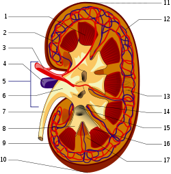
1. Renal pyramid • 2. Interlobular artery • 3. Renal artery • 4. Renal vein 5. Renal hilum • 6. Renal pelvis • 7. Ureter • 8. Minor calyx • 9. Renal capsule • 10. Inferior renal capsule • 11. Superior renal capsule • 12. Interlobular vein • 13. Nephron • 14. Minor calyx • 15. Major calyx • 16. Renal papilla • 17. Renal column The kidney has a bean-shaped structure; each kidney has a convex and concave surface. The concave surface, the renal hilum, is the point at which the renal artery enters the organ, and the renal vein and ureter leave. The kidney is surrounded by tough fibrous tissue, the renal capsule, which is itself surrounded by perinephric fat, renal fascia (of Gerota) and paranephric fat. The anterior (front) border of these tissues is the peritoneum, while the posterior (rear) border is the transversalis fascia.
The superior border of the right kidney is adjacent to the liver; and the spleen, for the left kidney. Therefore, both move down on inhalation.
The kidney is approximately 11–14 cm in length, 6 cm wide and 4 cm thick.
The substance, or parenchyma, of the kidney is divided into two major structures: superficial is the renal cortex and deep is the renal medulla. Grossly, these structures take the shape of 8 to 18 cone-shaped renal lobes, each containing renal cortex surrounding a portion of medulla called a renal pyramid (of Malpighi).[5] Between the renal pyramids are projections of cortex called renal columns (of Bertin). Nephrons, the urine-producing functional structures of the kidney, span the cortex and medulla. The initial filtering portion of a nephron is the renal corpuscle, located in the cortex, which is followed by a renal tubule that passes from the cortex deep into the medullary pyramids. Part of the renal cortex, a medullary ray is a collection of renal tubules that drain into a single collecting duct.
The tip, or papilla, of each pyramid empties urine into a minor calyx; minor calyces empty into major calyces, and major calyces empty into the renal pelvis, which becomes the ureter.
Blood supply
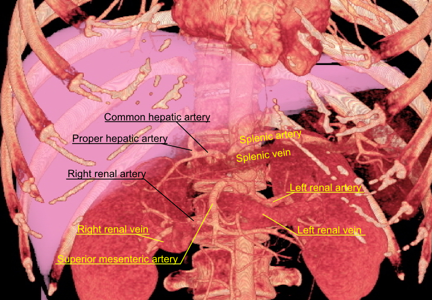
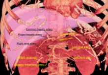 3D-rendered computed tomography, showing renal arteries and veins.
The kidneys receive blood from the renal arteries, left and right, which branch directly from the abdominal aorta. Despite their relatively small size, the kidneys receive approximately 20% of the cardiac output.[5]
Each renal artery branches into segmental arteries, dividing further into interlobar arteries which penetrate the renal capsule and extend through the renal columns between the renal pyramids. The interlobar arteries then supply blood to the arcuate arteries that run through the boundary of the cortex and the medulla. Each arcuate artery supplies several interlobular arteries that feed into the afferent arterioles that supply the glomeruli.
The interstitum (or interstitium) is the functional space in the kidney beneath the individual filters (glomeruli) which are rich in blood vessels. The interstitum absorbs fluid recovered from urine. Various conditions can lead to scarring and congestion of this area, which can cause kidney dysfunction and failure.
After filtration occurs the blood moves through a small network of venules that converge into interlobular veins. As with the arteriole distribution the veins follow the same pattern, the interlobular provide blood to the arcuate veins then back to the interlobar veins which come to form the renal vein exiting the kidney for transfusion for blood.
Histology
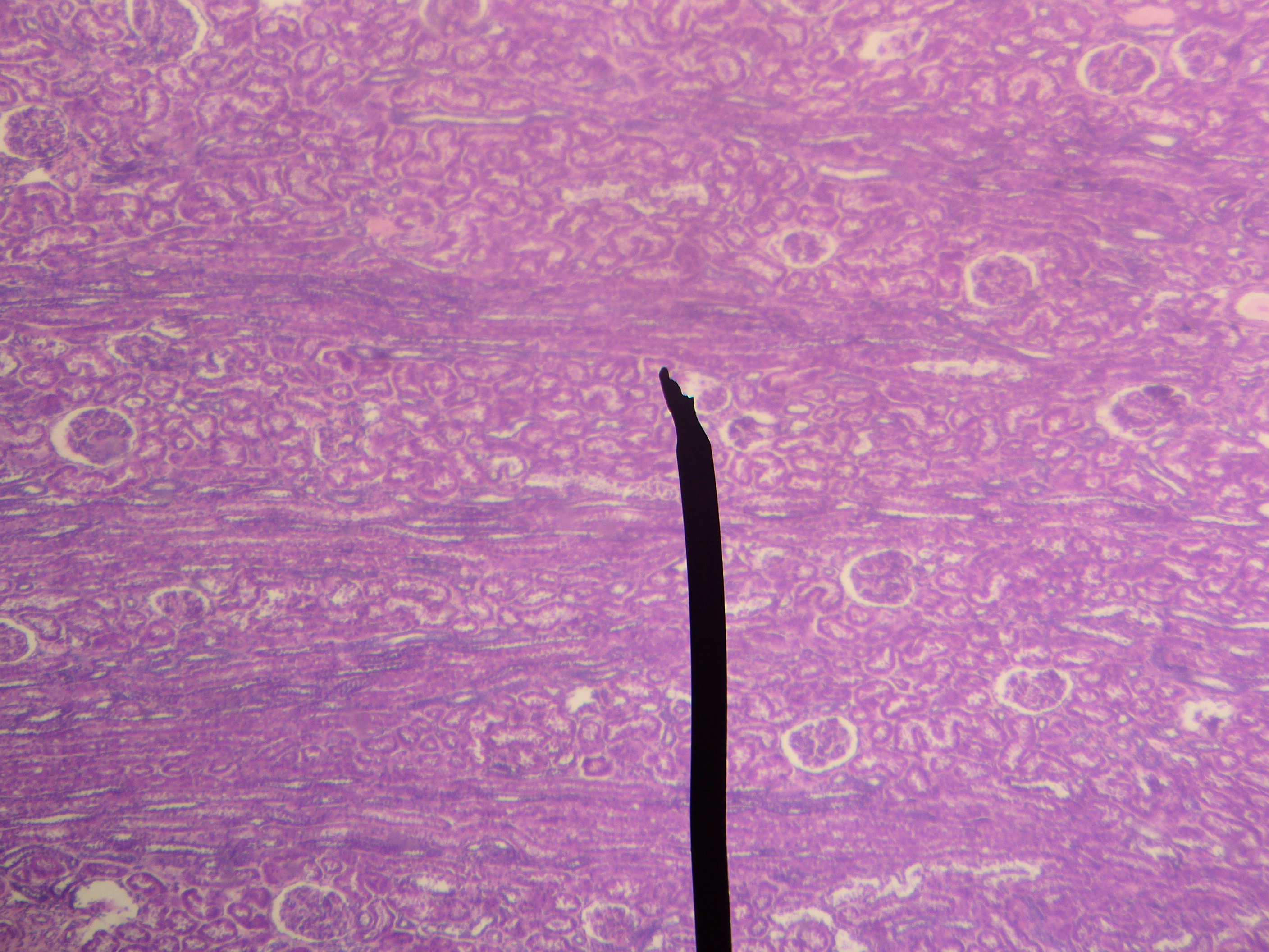
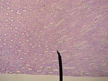 Microscopic photograph of the renal medulla
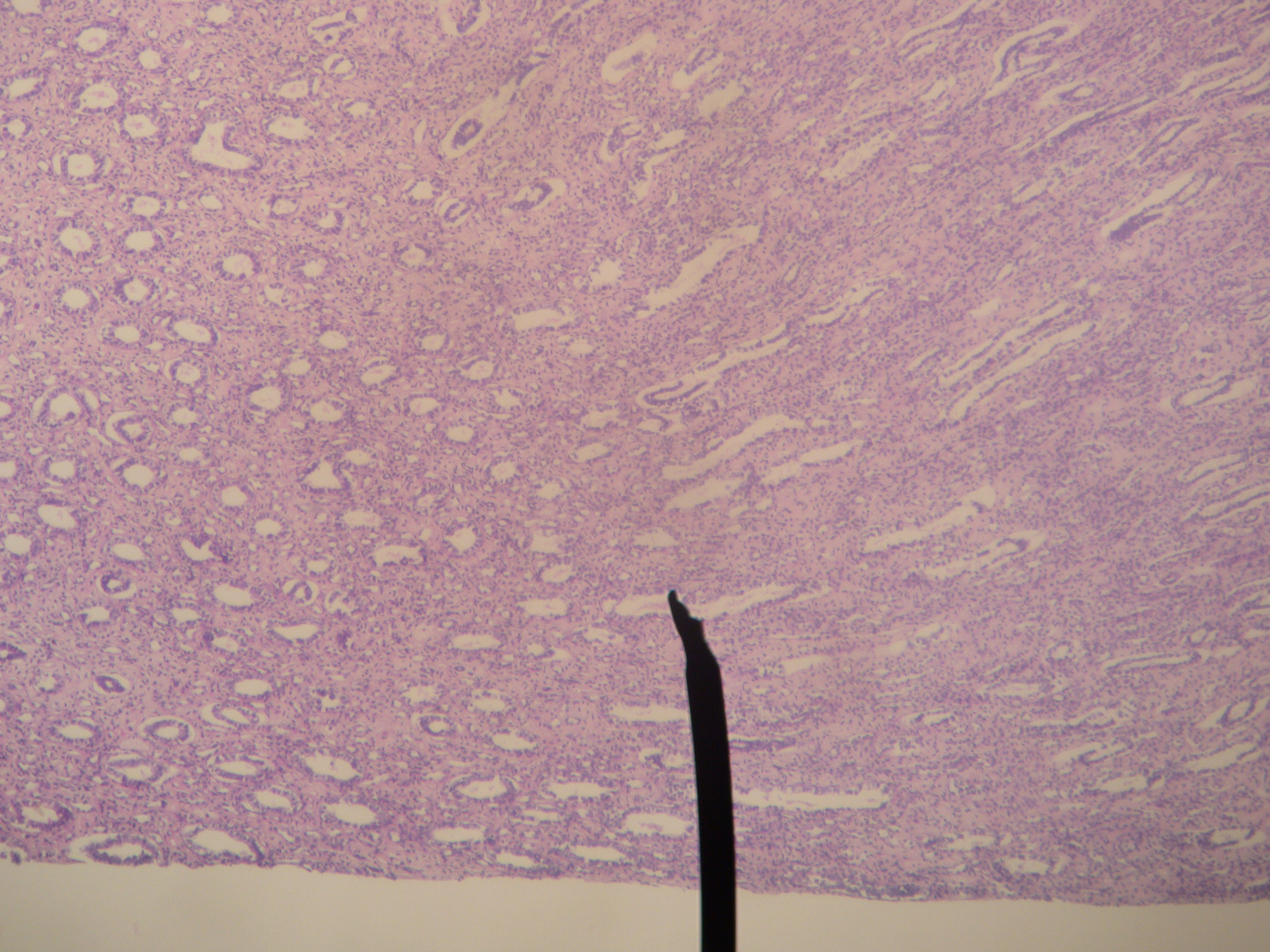
 Microscopic photograph of the renal cortex
Renal histology studies the structure of the kidney as viewed under a microscope. Various distinct cell types occur in the kidney, including:
- Kidney glomerulus parietal cell
- Kidney glomerulus podocyte
- Kidney proximal tubule brush border cell
- Loop of Henle thin segment cell
- Thick ascending limb cell
- Kidney distal tubule cell
- Kidney collecting duct cell
- Interstitial kidney cells
Innervation
The kidney and nervous system communicate via the renal plexus, whose fibers course along the renal arteries to reach the kidney.[7] Input from the sympathetic nervous system triggers vasoconstriction in the kidney, thereby reducing renal blood flow.[7] The kidney is not thought to receive input from the parasympathetic nervous system.[7] Sensory input from the kidney travels to the T10-11 levels of the spinal cord and is sensed in the corresponding dermatome.[7] Thus, pain in the flank region may be referred from the kidney.[7]
Functions
Main article: Renal physiologyThe kidney participates in whole-body homeostasis, regulating acid-base balance, electrolyte concentrations, extracellular fluid volume, and regulation of blood pressure. The kidney accomplishes these homeostatic functions both independently and in concert with other organs, particularly those of the endocrine system. Various endocrine hormones coordinate these endocrine functions; these include renin, angiotensin II, aldosterone, antidiuretic hormone, and atrial natriuretic peptide, among others.
Many of the kidney's functions are accomplished by relatively simple mechanisms of filtration, reabsorption, and secretion, which take place in the nephron. Filtration, which takes place at the renal corpuscle, is the process by which cells and large proteins are filtered from the blood to make an ultrafiltrate that eventually becomes urine. The kidney generates 180 liters of filtrate a day, while reabsorbing a large percentage, allowing for the generation of only approximately 2 liters of urine. Reabsorption is the transport of molecules from this ultrafiltrate and into the blood. Secretion is the reverse process, in which molecules are transported in the opposite direction, from the blood into the urine.
Excretion of wastes
The kidneys excrete a variety of waste products produced by metabolism. These include the nitrogenous wastes called "urea", from protein catabolism, as well as uric acid, from nucleic acid metabolism. Formation of urine is also the function of the kidney.
Acid-base homeostasis
Main article: Acid-base homeostasisTwo organ systems, the kidneys and lungs, maintain acid-base homeostasis, which is the maintenance of pH around a relatively stable value. The lungs contribute to acid-base homeostasis by regulating bicarbonate (HCO3-) concentration. The kidneys have two very important roles in maintaining the acid-base balance: to reabsorb bicarbonate from urine, and to excrete hydrogen ions into urine
Osmolality regulation
Any significant rise in plasma osmolality is detected by the hypothalamus, which communicates directly with the posterior pituitary gland. An increase in osmolality causes the gland to secrete antidiuretic hormone (ADH), resulting in water reabsorption by the kidney and an increase in urine concentration. The two factors work together to return the plasma osmolality to its normal levels.
ADH binds to principal cells in the collecting duct that translocate aquaporins to the membrane, allowing water to leave the normally impermeable membrane and be reabsorbed into the body by the vasa recta, thus increasing the plasma volume of the body.
There are two systems that create a hyperosmotic medulla and thus increase the body plasma volume: Urea recycling and the 'single effect.'
Urea is usually excreted as a waste product from the kidneys. However, when plasma blood volume is low and ADH is released the aquaporins that are opened are also permeable to urea. This allows urea to leave the collecting duct into the medulla creating a hyperosmotic solution that 'attracts' water. Urea can then re-enter the nephron and be excreted or recycled again depending on whether ADH is still present or not.
The 'Single effect' describes the fact that the ascending thick limb of the loop of Henle is not permeable to water but is permeable to NaCl. This allows for a countercurrent exchange system whereby the medulla becomes increasingly concentrated, but at the same time setting up an osmotic gradient for water to follow should the aquaporins of the collecting duct be opened by ADH.
Blood pressure regulation
Main articles: Blood pressure regulation and Renin-angiotensin systemLong-term regulation of blood pressure predominantly depends upon the kidney. This primarily occurs through maintenance of the extracellular fluid compartment, the size of which depends on the plasma sodium concentration. Although the kidney cannot directly sense blood pressure, changes in the delivery of sodium and chloride to the distal part of the nephron alter the kidney's secretion of the enzyme renin. When the extracellular fluid compartment is expanded and blood pressure is high, the delivery of these ions is increased and renin secretion is decreased. Similarly, when the extracellular fluid compartment is contracted and blood pressure is low, sodium and chloride delivery is decreased and renin secretion is increased in response.
Renin is the first in a series of important chemical messengers that comprise the renin-angiotensin system. Changes in renin ultimately alter the output of this system, principally the hormones angiotensin II and aldosterone. Each hormone acts via multiple mechanisms, but both increase the kidney's absorption of sodium chloride, thereby expanding the extracellular fluid compartment and raising blood pressure. When renin levels are elevated, the concentrations of angiotensin II and aldosterone increase, leading to increased sodium chloride reabsorption, expansion of the extracellular fluid compartment, and an increase in blood pressure. Conversely, when renin levels are low, angiotensin II and aldosterone levels decrease, contracting the extracellular fluid compartment, and decreasing blood pressure.
Hormone secretion
The kidneys secrete a variety of hormones, including erythropoietin, and the enzyme renin. Erythropoietin is released in response to hypoxia (low levels of oxygen at tissue level) in the renal circulation. It stimulates erythropoiesis (production of red blood cells) in the bone marrow. Calcitriol, the activated form of vitamin D, promotes intestinal absorption of calcium and the renal reabsorption of phosphate. Part of the renin-angiotensin-aldosterone system, renin is an enzyme involved in the regulation of aldosterone levels.
Development
Main article: Kidney developmentThe mammalian kidney develops from intermediate mesoderm. Kidney development, also called nephrogenesis, proceeds through a series of three successive phases, each marked by the development of a more advanced pair of kidneys: the pronephros, mesonephros, and metanephros.[8]
Evolutionary adaptation
Kidneys of various animals show evidence of evolutionary adaptation and have long been studied in ecophysiology and comparative physiology. Kidney morphology, often indexed as the relative medullary thickness, is associated with habitat aridity among species of mammals.[9]
Etymology
Medical terms related to the kidneys commonly use terms such as renal and the prefix nephro-. The adjective renal, meaning related to the kidney, is from the Latin rēnēs, meaning kidneys; the prefix nephro- is from the Ancient Greek word for kidney, nephros (νεφρός).[10] For example, surgical removal of the kidney is a nephrectomy, while a reduction in kidney function is called renal dysfunction.
Diseases and disorders
Main article: NephropathyCongenital
- Congenital hydronephrosis
- Congenital obstruction of urinary tract
- Duplex kidneys, or double kidneys, occur in approximately 1% of the population. This occurrence normally causes no complications, but can occasionally cause urine infections.[11][12]
- Duplicated ureter occurs in approximately one in 100 live births
- Horseshoe kidney occurs in approximately one in 400 live births
- Polycystic kidney disease
- Autosomal dominant polycystic kidney disease afflicts patients later in life. Approximately one in 1000 people will develop this condition
- Autosomal recessive polycystic kidney disease is far less common, but more severe, than the dominant condition. It is apparent in utero or at birth.
- Renal agenesis. Failure of one kidney to form occurs in approximately one in 750 live births. Failure of both kidneys to form is invariably fatal.
- Renal dysplasia
- Unilateral small kidney
- Multicystic dysplastic kidney occurs in approximately one in every 2400 live births
- Ureteropelvic Junction Obstruction or UPJO; although most cases appear congenital, some appear to be an acquired condition[13]
Acquired
 Drawing of an enlarged kidney by John Hunter.
- Diabetic nephropathy
- Glomerulonephritis
- Hydronephrosis is the enlargement of one or both of the kidneys caused by obstruction of the flow of urine.
- Interstitial nephritis
- Kidney stones (nephrolithiasis) are a relatively common and particularly painful disorder.
- Kidney tumors
- Wilms tumor
- Renal cell carcinoma
- Lupus nephritis
- Minimal change disease
- In nephrotic syndrome, the glomerulus has been damaged so that a large amount of protein in the blood enters the urine. Other frequent features of the nephrotic syndrome include swelling, low serum albumin, and high cholesterol.
- Pyelonephritis is infection of the kidneys and is frequently caused by complication of a urinary tract infection.
- Renal failure
Kidney Failure
Main article: Renal failureGenerally, humans can live normally with just one kidney, as one has more functioning renal tissue than is needed to survive. Only when the amount of functioning kidney tissue is greatly diminished does one develop chronic kidney disease. Renal replacement therapy, in the form of dialysis or kidney transplantation, is indicated when the glomerular filtration rate has fallen very low or if the renal dysfunction leads to severe symptoms.
In other animals
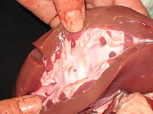 A pig's kidney opened.
In the majority of vertebrates, the mesonephros persists into the adult, albeit usually fused with the more advanced metanephros; only in amniotes is the mesonephros restricted to the embryo. The kidneys of fish and amphibians are typically narrow, elongated organs, occupying a significant portion of the trunk. The collecting ducts from each cluster of nephrons usually drain into an archinephric duct, which is homologous with the vas deferens of amniotes. However, the situation is not always so simple; in cartilaginous fish and some amphibians, there is also a shorter duct, similar to the amniote ureter, which drains the posterior (metanephric) parts of the kidney, and joins with the archinephric duct at the bladder or cloaca. Indeed, in many cartilaginous fish, the anterior portion of the kidney may degenerate or cease to function altogether in the adult.[14]
In the most primitive vertebrates, the hagfish and lampreys, the kidney is unusually simple: it consists of a row of nephrons, each emptying directly into the archinephric duct. Invertebrates may possess excretory organs that are sometimes referred to as "kidneys", but, even in Amphioxus, these are never homologous with the kidneys of vertebrates, and are more accurately referred to by other names, such as nephridia.[14]
The kidneys of reptiles consist of a number of lobules arranged in a broadly linear pattern. Each lobule contains a single branch of the ureter in its centre, into which the collecting ducts empty. Reptiles have relatively few nephrons compared with other amniotes of a similar size, possibly because of their lower metabolic rate.[14]
Birds have relatively large, elongated kidneys, each of which is divided into three or more distinct lobes. The lobes consists of several small, irregularly arranged, lobules, each centred on a branch of the ureter. Birds have small glomeruli, but about twice as many nephrons as similarly sized mammals.[14]
The human kidney is fairly typical of that of mammals. Distinctive features of the mammalian kidney, in comparison with that of other vertebrates, include the presence of the renal pelvis and renal pyramids, and of a clearly distinguishable cortex and medulla. The latter feature is due to the presence of elongated loops of Henle; these are much shorter in birds, and not truly present in other vertebrates (although the nephron often has a short intermediate segment between the convoluted tubules). It is only in mammals that the kidney takes on its classical "kidney" shape, although there are some exceptions, such as the multilobed reniculate kidneys of cetaceans.[14]
History
The Latin term renes is related to the English word "reins", a synonym for the kidneys in Shakespearean English (e.g. Merry Wives of Windsor 3.5), which was also the time the King James Version was translated. Kidneys were once popularly regarded as the seat of the conscience and reflection,[15][16] and a number of verses in the Bible (e.g. Ps. 7:9, Rev. 2:23) state that God searches out and inspects the kidneys, or "reins", of humans. Similarly, the Talmud (Berakhoth 61.a) states that one of the two kidneys counsels what is good, and the other evil.
Animal kidneys as food
 Hökarpanna, Swedish pork and kidney stew
The kidneys of animals can be cooked and eaten by humans (along with other offal).
Kidneys are usually grilled or sautéed, but in more complex dishes they are stewed with a sauce that will improve their flavor. In many preparations, kidneys are combined with pieces of meat or liver, as in mixed grill or meurav Yerushalmi. Among the most reputed kidney dishes, the British steak and kidney pie, the Swedish hökarpanna (pork and kidney stew), the French rognons de veau sauce moutarde (veal kidneys in mustard sauce) and the Spanish riñones al Jerez (kidneys stewed in sherry sauce), deserve special mention.[17]
See also
References
- ^ Cotran, RS S.; Kumar, Vinay; Fausto, Nelson; Robbins, Stanley L.; Abbas, Abul K. (2005). Robbins and Cotran pathologic basis of disease. St. Louis, MO: Elsevier Saunders. ISBN 0-7216-0187-1.
- ^ "Kidneys Location Stock Illustration". http://www.indexedvisuals.com/scripts/ivstock/pic.asp?id=118-100.
- ^ [1][dead link]
- ^ Bålens ytanatomy (Superficial anatomy of the trunk). Anca Dragomir, Mats Hjortberg and Godfried M. Romans. Section for human anatomy at the Department of medical cell biology, Uppsala university, Sweden.
- ^ a b c Walter F., PhD. Boron (2004). Medical Physiology: A Cellular And Molecular Approach. Elsevier/Saunders. ISBN 1-4160-2328-3.
- ^ Glodny B, Unterholzner V, Taferner B, et al. (2009). "Normal kidney size and its influencing factors - a 64-slice MDCT study of 1.040 asymptomatic patients". BMC Urology 9: 19. doi:10.1186/1471-2490-9-19. PMC 2813848. PMID 20030823. http://www.biomedcentral.com/1471-2490/9/19.
- ^ a b c d e Bard, Johnathan; Vize, Peter D.; Woolf, Adrian S. (2003). The kidney: from normal development to congenital disease. Boston: Academic Press. p. 154. ISBN 0-12-722441-6. http://books.google.com/?id=ctOm-cPwo60C&pg=PA154.
- ^ Bruce M. Carlson (2004). Human Embryology and Developmental Biology (3rd ed.). Saint Louis: Mosby. ISBN 0-323-03649-X.
- ^ Al-kahtani, M. A.; C. Zuleta, E. Caviedes-Vidal, and T. Garland, Jr. (2004). "Kidney mass and relative medullary thickness of rodents in relation to habitat, body size, and phylogeny". Physiological and Biochemical Zoology 77 (3): 346–365. doi:10.1086/420941. PMID 15286910. http://www.biology.ucr.edu/people/faculty/Garland/Al-kahtaniEA2004.pdf.
- ^ Maton, Anthea; Jean Hopkins, Charles William McLaughlin, Susan Johnson, Maryanna Quon Warner, David LaHart, Jill D. Wright (1993). Human Biology and Health. Englewood Cliffs, New Jersey, USA: Prentice Hall. ISBN 0-13-981176-1.
- ^ Sample, Ian (2008-02-19). "How many people have four kidneys?". The Guardian (London). http://www.guardian.co.uk/society/2008/feb/19/health.
- ^ "Girl's Kidneys Fail, But Doctors Find Double Valves, Saving Her Life". Abcnews.go.com. 2010-05-18. http://abcnews.go.com/Health/girls-kidneys-fail-doctors-find-double-valves-saving/story?id=10668525. Retrieved 2011-01-03.
- ^ Stephen Jones, J.; Inderbir S. Gill, Raymond Rackley (2006). Operative Urology at the Cleveland Clinic. Andrew C. Novick, Inderbir S. Gill, Eric A. Klein, Jonathan H. Ross (eds.). Totowa, NJ: Humana Press. ISBN 978-1-58829-081-6. http://www.springerlink.com/content/p483835241230k88/. Retrieved 2010-10-09.
- ^ a b c d e Romer, Alfred Sherwood; Parsons, Thomas S. (1977). The Vertebrate Body. Philadelphia, PA: Holt-Saunders International. pp. 367–376. ISBN 0-03-910284-X.
- ^ The Patient as Person: Explorations in Medical Ethics p. 60 by Paul Ramsey, Margaret Farley, Albert Jonsen, William F. May (2002)
- ^ History of Nephrology 2 p. 235 by International Association for the History of Nephrology Congress, Garabed Eknoyan, Spyros G. Marketos, Natale G. De Santo - 1997; Reprint of American Journal of Nephrology; v. 14, no. 4-6, 1994.
- ^ Rognons dans les recettes (French)
External links
- The NephCure Foundation offers educational materials on the kidney diseases/conditions Nephrotic Syndrome and FSGS
- The Kidney Foundation of Canada
- electron microscopic images of the kidney (Dr. Jastrow's EM-Atlas)
- European Renal Genome project kidney function tutorial
- Kidney Foundation of Canada kidney disease information
- Renal Fellow Network: Structure & Function of Other Animals' Kidneys
- Kidney Stones
- Kidney Diseases
- Kidney Information
- Animated Presentatin on Kidney Function
- CAT Scans of various kidney diseases and conditions - CT Cases
Human systems and organs TA 2–4:
MSBone (Carpus · Collar bone (clavicle) · Thigh bone (femur) · Fibula · Humerus · Mandible · Metacarpus · Metatarsus · Ossicles · Patella · Phalanges · Radius · Skull (cranium) · Tarsus · Tibia · Ulna · Rib · Vertebra · Pelvis · Sternum) · CartilageTA 5–11:
splanchnic/
viscusmostly
Thoracicmostly
AbdominopelvicDigestive system+
adnexaMouth (Salivary gland, Tongue) · upper GI (Oropharynx, Laryngopharynx, Esophagus, Stomach) · lower GI (Small intestine, Appendix, Colon, Rectum, Anus) · accessory (Liver, Biliary tract, Pancreas)TA 12–16 Blood
(Non-TA)General anatomy: systems and organs, regional anatomy, planes and lines, superficial axial anatomy, superficial anatomy of limbsAnatomy: urinary system (TA A08, TH H3.06, GA 11.1215) Abdomen KidneysLayersRenal tubuleFiltrationPelvis Apex • Uvula • Neck • Median umbilical ligament • Muscular layer (Trigone • Detrusor) • Mucosa • SubmucosaCategories:


Wikimedia Foundation. 2010.