- Major calyx
-
Major calyx 
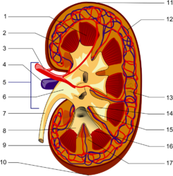
1. Renal triangle
2. Efferent artery
3. Renal artery
4. Renal vein
5. Renal hilum
6. Renal pelvis
7. Ureter
8. Minor calyx
9. Renal capsule10. Inferior renal capsule
11. Superior renal capsule
12. Afferent vein
13. Nephron
14. Minor calyx
15. Major calyx
16. Renal papilla
17. Renal column
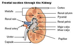
Frontal section through the kidney Latin calices renales majores Gray's subject #254 1225 Precursor Ureteric bud MeSH Kidney+calices Two or three minor calyces converge to form a major calyx. The major calyx, in the kidney, surrounds the apex of the renal pyramids. Urine formed in the kidney passes through a renal papilla at the apex into a minor calyx then into major calyx before passing through the renal pelvis into the ureter.
Peristalsis of the smooth muscle originating in pace-maker cells originating in the walls of the calyces propels urine through the renal pelvis and ureters to the bladder.
Additional images
See also
External links
- SUNY Labs 40:06-0109 - "Posterior Abdominal Wall: Internal Structure of a Kidney"
- Histology at BU 15901loa - "Urinary System: neonatal kidney"
- posteriorabdomen at The Anatomy Lesson by Wesley Norman (Georgetown University) (renalpelvis)
- Diagram at bway.net
Anatomy: urinary system (TA A08, TH H3.06, GA 11.1215) Abdomen LayersRenal tubuleFiltrationPelvis Apex • Uvula • Neck • Median umbilical ligament • Muscular layer (Trigone • Detrusor) • Mucosa • SubmucosaCategories:- Genitourinary system stubs
- Kidney anatomy
Wikimedia Foundation. 2010.


