- Renal medulla
Infobox Anatomy
Name = PAGENAME
Latin =
GraySubject = 253
GrayPage = 1221
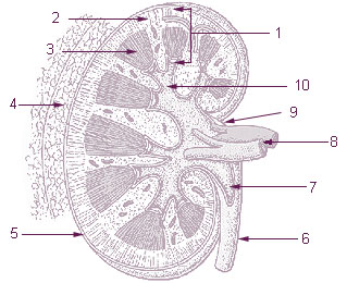
Caption = 1:Parenchyma
2: Cortex
3: Medulla
4:Perirenal fat
5: Capsule
6:Ureter
7: Pelvis of kidney
8:Renal artery andRenal vein
9: Hilus
10: Calyx
Caption2 = Vertical section of kidney. (Label "medullary sub." visible near top.)
System =
MeshName = Kidney+medulla
MeshNumber = A05.810.453.466
DorlandsPre =
DorlandsSuf =
The renal medulla is the innermost part of thekidney . The renal medulla is split up into a number of sections, known as therenal pyramids . Blood enters into the kidney via the renal artery, which then splits up to form the arcuate arterioles. Thearcuate arterioles each in turn branch into interlobular arterioles, which finally reach the glomeruli. At the glomerulus the blood reaches a highly disfavourable pressure gradient and a large exchange surface area, which forces theserum portion of the blood out of the vessel into the renal tubules. Flow continues through the renal tubules, including the proximal tubule, theLoop of Henle , and finally leaves the kidney by means of thecollecting duct , leading to the renalureter .
=AdditionalExternal links
* [http://www.medicalengineer.co.uk/Microcirculation+of+the+Renal+Medulla.php Medical Engineer - Renal Microcirculation]
* [http://education.vetmed.vt.edu/Curriculum/VM8054/Labs/Lab23/Examples/exmedulla.htm VetMed - Renal Medulla]
Wikimedia Foundation. 2010.