- Renal cortex
-
For other uses, see Cortex (disambiguation).
Renal cortex 
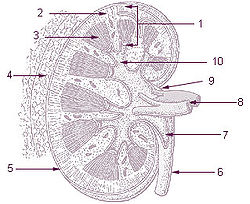
1: Parenchyma
2: Cortex
3: Medulla
4: Perirenal fat
5: Capsule
6: Ureter7: Pelvis of kidney
8: Renal artery and Renal vein
9: Hilus
10: Calyx
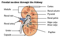
Frontal section through the kidney Gray's subject #253 1221 MeSH Kidney+cortex The renal cortex is the outer portion of the kidney between the renal capsule and the renal medulla. In the adult, it forms a continuous smooth outer zone with a number of projections (cortical columns) that extend down between the pyramids. It contains the renal corpuscles and the renal tubules except for parts of the loop of Henle which descend into the renal medulla. It also contains blood vessels and cortical collecting ducts.
The renal cortex is the part of the kidney where ultrafiltration occurs. Erythropoietin is produced in the renal cortex.
Additional images
-
CD10 immunohistochemical staining of normal kidney. CD10 stains the proximal convoluted tubules and glomeruli.
Contains afferent arterioles
External links
- SUNY Labs 40:06-0103 - "Posterior Abdominal Wall: Internal Structure of a Kidney"
Anatomy: urinary system (TA A08, TH H3.06, GA 11.1215) Abdomen LayersRenal fascia • Renal capsule • Renal cortex (Renal column) • Renal medulla (Renal sinus, Renal pyramids, medullary interstitium) • Renal lobe • Cortical lobule • Medullary ray • NephronRenal tubuleFiltrationPelvis Apex • Uvula • Neck • Median umbilical ligament • Muscular layer (Trigone • Detrusor) • Mucosa • SubmucosaCategories:- Kidney anatomy
- Genitourinary system stubs
Wikimedia Foundation. 2010.


