- Thick ascending limb of loop of Henle
-
Thick ascending limb of loop of Henle 
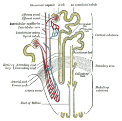
Scheme of renal tubule and its vascular supply. (Labeled at center left.) 
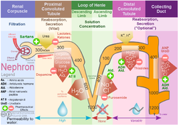
Nephron ion flow diagram Latin tubulus rectus distalis Gray's subject #253 1223 The thick ascending limb of loop of Henle (TAL) also known as distal straight tubule, is a segment of the nephron in the kidney. It can be divided into two parts: that in the renal medulla, and that in the renal cortex.
Medullary thick ascending limb
The medullary thick ascending limb remains impermeable to water. Sodium, potassium (K+) and chloride (Cl-) ions are reabsorbed by active transport. K+ is passively transported along its concentration gradient through a K+ leak channel in the apical aspect of the cells, back into the lumen of the ascending limb. This K+ "leak" generates a positive electrochemical potential difference in the lumen. The electrical gradient drives more reabsorption of Na+, as well as other cations such as magnesium (Mg2+) and importantly calcium Ca2+.
Loop diuretics such as furosemide block the K+/Na+/2Cl- co-transporter.
This is also the part of the tubule that generates Tamm-Horsfall protein. The function of this protein is not well understood, but is responsible for creating urinary casts.
Cortical thick ascending limb
The difference between the medullary and cortical thick ascending limbs is mainly anatomical. Functionally, they are very similar.
The cortical thick ascending limb drains urine into the distal convoluted tubule.
External links
This article was originally based on an entry from a public domain edition of Gray's Anatomy. As such, some of the information contained within it may be outdated.
Anatomy: urinary system (TA A08, TH H3.06, GA 11.1215) Abdomen LayersRenal tubuleRenal corpuscle (Glomerulus, Bowman's capsule) → Proximal tubule → Loop of Henle (Descending, Thin ascending, Thick ascending) → Distal convoluted tubule → Connecting tubule → Collecting ducts aka Duct of Belini → Renal papilla → Minor calyx → Major calyx → Renal pelvisFiltrationPelvis Apex • Uvula • Neck • Median umbilical ligament • Muscular layer (Trigone • Detrusor) • Mucosa • SubmucosaCategories:- Genitourinary system stubs
- Kidney anatomy
Wikimedia Foundation. 2010.
