- Embryology
-
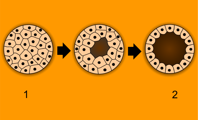
 1 - morula, 2 - blastula
 1 - blastula, 2 - gastrula with blastopore; orange - ectoderm, red - endoderm.
Embryology (from Greek ἔμβρυον, embryon, "unborn, embryo"; and -λογία, -logia) is a science which is about the development of an embryo from the fertilization of the ovum to the fetus stage. After cleavage, the dividing cells, or morula, becomes a hollow ball, or blastula, which develops a hole or pore at one end.
In bilateral animals, the blastula develops in one of two ways that divides the whole animal kingdom into two halves (see: Embryological origins of the mouth and anus). If in the blastula the first pore (blastopore) becomes the mouth of the animal, it is a protostome; if the first pore becomes the anus then it is a deuterostome. The protostomes include most invertebrate animals, such as insects, worms and molluscs, while the deuterostomes include the vertebrates. In due course, the blastula changes into a more differentiated structure called the gastrula.
The gastrula with its blastopore soon develops three distinct layers of cells (the germ layers) from which all the bodily organs and tissues then develop:
- The innermost layer, or endoderm, gives rise to the digestive organs, lungs and bladder.
- The middle layer, or mesoderm, gives rise to the muscles, skeleton and blood system.
- The outer layer of cells, or ectoderm, gives rise to the nervous system and skin.
In humans, the term embryo refers to the ball of dividing cells from the moment the zygote implants itself in the uterus wall until the end of the eighth week after conception. Beyond the eighth week, the developing human is then called a fetus. Embryos in many species often appear similar to one another in early developmental stages. The reason for this similarity is because species have a shared evolutionary history. These similarities among species are called homologous structures, which are structures that have the same or similar function and mechanism having evolved from a common ancestor.
Contents
History
 Human embryo at six weeks gestational age
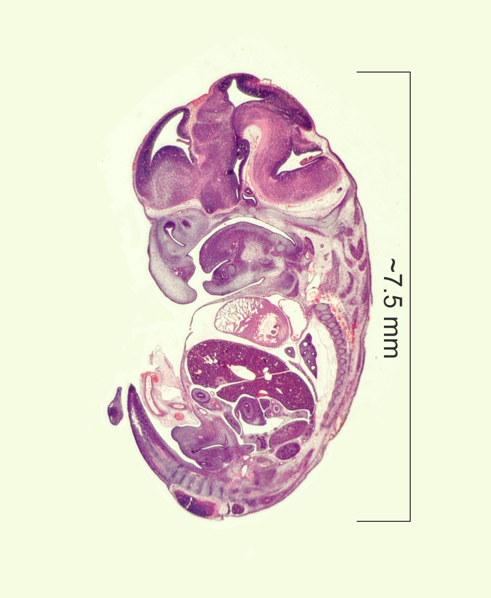
 Histological film 10 day mouse embryo
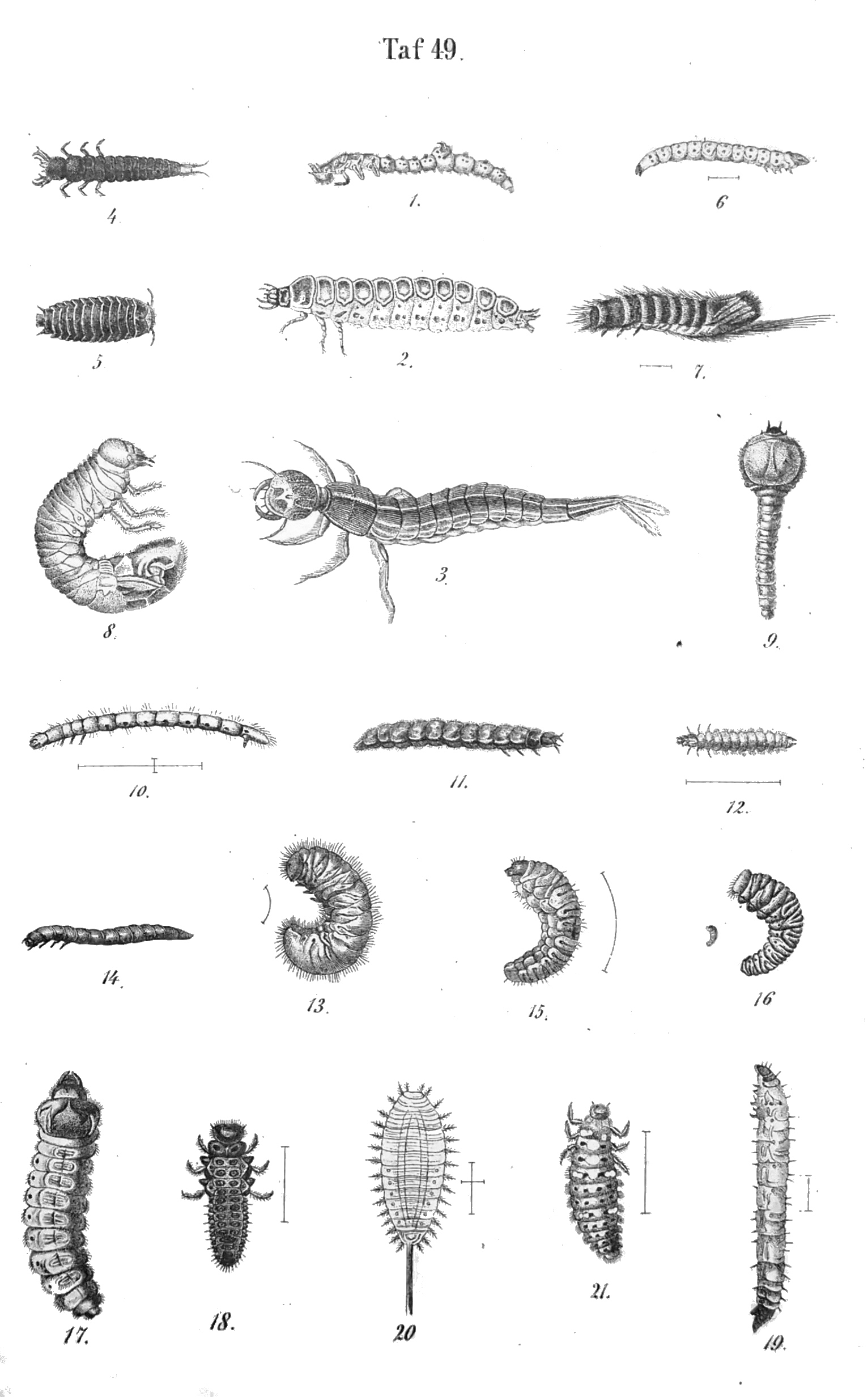
 Beetle larvae
As recently as the 18th century, the prevailing notion in human embryology was preformation: the idea that semen contains an embryo — a preformed, miniature infant, or "homunculus" — that simply becomes larger during development. The competing explanation of embryonic development was epigenesis, originally proposed 2,000 years earlier by Aristotle. Much early embryology came from the work of the great Italian anatomists: Aldrovandi, Aranzio, Leonardo da Vinci, Marcello Malpighi, Gabriele Falloppio, Girolamo Cardano, Emilio Parisano, Fortunio Liceti, Stefano Lorenzini, Spallanzani, Enrico Sertoli, Mauro Rusconi, etc.[1] According to epigenesis, the form of an animal emerges gradually from a relatively formless egg. As microscopy improved during the 19th century, biologists could see that embryos took shape in a series of progressive steps, and epigenesis displaced preformation as the favored explanation among embryologists.[2]
Finally Karl Ernst von Baer discovered the mammalian ovum in 1827.[3][4][5] Modern embryological pioneers include Charles Darwin, Ernst Haeckel, J.B.S. Haldane, and Joseph Needham. Other important contributors include William Harvey, Kaspar Friedrich Wolff, Heinz Christian Pander, August Weismann, Gavin de Beer, Ernest Everett Just, and Edward B. Lewis.
After the 1950s, with the DNA helical structure being unravelled and the increasing knowledge in the field of molecular biology, developmental biology emerged as a field of study which attempts to correlate the genes with morphological change, and so tries to determine which genes are responsible for each morphological change that takes place in an embryo, and how these genes are regulated.
Vertebrate and invertebrate embryology
Many principles of embryology apply to both invertebrate animals as well as to vertebrates.[6] Therefore, the study of invertebrate embryology has advanced the study of vertebrate embryology. However, there are many differences as well. For example, numerous invertebrate species release a larva before development is complete; at the end of the larval period, an animal for the first time comes to resemble an adult similar to its parent or parents. Although invertebrate embryology is similar in some ways for different invertebrate animals, there are also countless variations. For instance, while spiders proceed directly from egg to adult form many insects develop through at least one larval stage
Modern embryology research
Currently, embryology has become an important research area for studying the genetic control of the development process (e.g. morphogens), its link to cell signalling, its importance for the study of certain diseases and mutations and in links to stem cell research.
See also
- Ontogeny
- Embryogenesis
- Recapitulation theory
- Prenatal development
- Protostomes
- Deuterostomes
- Germ layers
- Epigenesis (biology)
- Developmental biology
- Cell signalling
- Hedgehog signaling pathway
- Morphogens
- French flag model
- Embryo drawing
References
- ^ Massimo De Felici, Gregorio Siracus, The rise of embryology in Italy: from the Renaissance to the early 20th Century, Int. J. Dev. Biol. 44: 515-521 (2000).
- ^ Campbell et al. (p. 987)
- ^ K. J. Betteridge (1981). "An historical look at embryo transfer". Reproduction. The Journal of the Society for Reproduction and Fertility 62: 1–13. http://www.reproduction-online.org/content/62/1/1.full.pdf+html. "Three years later, the Estonian, Karl Ernst von Baer, finally found the true mammalian egg in a pet dog (von Baer, 1827)."
- ^ Lois N. Magner (2005). History of the Life Sciences. New York. Basel: Marcel Dekker. p. 166. http://books.google.com/books?id=EnG0gshuCBAC&printsec=frontcover&dq=History+of+the+Life+Sciences&hl=et&ei=6feXTp_PG-_44QSOkJCZBA&sa=X&oi=book_result&ct=result&resnum=1&ved=0CC0Q6AEwAA#v=onepage&q&f=false.
- ^ Alex Lopata (2009). "History of the Egg in Embryology". Journal of Mammalian Ova Research 26: 2–9. http://www.bioone.org/doi/full/10.1274/jmor.26.2.
- ^ Parker, Sybil. "Invertebrate Embryology," McGraw-Hill Encyclopedia of Science & Technology (McGraw-Hill 1997).
Embryology - History of embryology as a science." Science Encyclopedia. Web. 06 Nov. 2009. <http://science.jrank.org/pages/2452/Embryology.html>.
"Germ layer." Encyclopædia Britannica. 2009. Encyclopædia Britannica Online. 06 Nov. 2009 <http://www.britannica.com/EBchecked/topic/230597/germ-layer>.Further reading
- Apostoli, Pietro; Catalani, Simona (2011). "Chapter 11. Metal Ions Affecting Reproduction and Development". In Astrid Sigel, Helmut Sigel and Roland K. O. Sigel. Metal Ions in Toxicology. Metal Ions in Life Sciences. 8. RSC Publishing. pp. 263–303. doi:10.1039/9781849732116-00263.
- Scott F. Gilbert. Developmental Biology. Sinauer, 2003. ISBN 0-87893-258-5.
- Lewis Wolpert. Principles of Development. Oxford University Press, 2006. ISBN 0-19-927536-X.
External links
- Indiana University's Human Embryology Animations
- What is a human admixed embryo?
- UNSW Embryology | UNSW Embryology Large resource of information and media
- Definition of embryo according to Webster
Developmental biology > Human embryogenesis (development of embryo) and development of fetus (TE E2.0) First three
weeksWeek 1Fertilization · Oocyte activation · Zygote · Cleavage · Morula · Blastula (Blastomere) · Blastocyst · Inner cell massWeek 2
(Bilaminar)Week 3
(Trilaminar)Archenteron/Primitive streak (Primitive pit, Primitive knot/Blastopore, Primitive groove) · Gastrula/Gastrulation · Regional specification · Embryonic discSplanchnopleuric mesenchymeChorda- · Paraxial (Somite/Somitomere) · Intermediate · Lateral plate (Intraembryonic coelom, Splanchnopleuric mesenchyme/Somatopleuric mesenchyme)Human development of head and neck (GA 1.65, TE E5.3, 5.4) Face Nasal placode · Nasal pit · nasal prominences (Lateral, Medial) · Intermaxillary segment
Frontonasal prominence · Maxillary prominence · Mandibular prominence (Meckel's cartilage)Oral cavity Primary palate · Secondary palateLateral lingual swelling · Tuberculum impar · Copula linguae · Hypopharyngeal eminence · Gustatory placodeGeneral
branchial apparatusEmbryology of bones, joints, and muscles (GA 2.80, TE E5.0-2) Ossification Lower limbHeadcranium: Ossification of occipital bone · Ossification of frontal bone · Ossification of temporal bone · Ossification of sphenoid · Ossification of ethmoid
facial bones: Ossification of vomer (Sutura vomerina · Foramen vomerinum · Meatus vomerinus · Fissura vomerina) · Ossification of maxilla · Ossification of mandibleOtherOther M: JNT
anat(h/c, u, t, l)/phys
noco(arth/defr/back/soft)/cong, sysi/epon, injr
proc, drug(M01C, M4)
Prenatal development/Mammalian development of circulatory system (GA 5, TE E5.11) Heart development Tubular heartSepta/ostiaAtrioventricular cushions/Septum intermedium · Primary interatrial foramen · Septum primum (Foramen secundum) · Septum secundum (Foramen ovale) · Aorticopulmonary septumOtherAtrioventricular canal · Primary interventricular foramenVasculogenesis,
angiogenesis,
and lymphangiogenesisBlood island of umbilical vesicle
Development of arteriesDevelopment of veinsDevelopment of lymph vesselsLymph sacsDevelopment of circulatory system about teeth near childrenanuli: Anulus sanguineus perienameleus · lacunae: Lacuna sanguinea supraenamelea (Ductus sanguineus mesialis · Ductus sanguineus distalis · Ductus sanguineus lingualis · Ductus sanguineus palatinus · Ductus sanguineus buccalis · Ductus sanguineus labialis), Lacuna sanguinea apicalis, Lacuna sanguinea periodontalis, Lacuna sanguinea parodontalis, Lacuna sanguinea gingivalisExtraembryonic
hemangiogenesisFetal circulation Prenatal development/Mammalian development of nervous system (GA 9.733 and GA 10.1002, TE E5.13-16) Neurogenesis Cranial neural crest (Cardiac neural crest complex) · Truncal neural crestRostral neuropore
Cephalic flexure · Pontine flexure
Alar plate (sensory) · Basal plate (motor)
Germinal matrixEye development Auditory development M: EYE
anat(g/a/p)/phys/devp/prot
noco/cong/tumr, epon
proc, drug(S1A/1E/1F/1L)
M: EAR
anat(e/p)/phys/devp
noco/cong, epon
proc, drug(S2)
Prenatal development/Mammalian development of nervous system (GA 9.733 and GA 10.1002, TE E5.13-16) Neurogenesis Cranial neural crest (Cardiac neural crest complex) · Truncal neural crestRostral neuropore
Cephalic flexure · Pontine flexure
Alar plate (sensory) · Basal plate (motor)
Germinal matrixEye development Auditory development M: EYE
anat(g/a/p)/phys/devp/prot
noco/cong/tumr, epon
proc, drug(S1A/1E/1F/1L)
M: EAR
anat(e/p)/phys/devp
noco/cong, epon
proc, drug(S2)
Prenatal development/Mammalian development of respiratory system (overview) (GA 11.1071, TE E5.5) Upper Lower Laryngotracheal groove · Respiratory budPrenatal development/Mammalian development of digestive system, coelom and septa, and mesenchymal mesenteric masses (GA 11.1101, TE E5.4, 5.8-9) Gut Upper GI tract and accessoryForegut: upper GI (Buccopharyngeal membrane, Rathke's pouch, Tracheoesophageal septum) · accessory (Pancreatic bud, Hepatic diverticulum)Abdominopelvic OtherIntra-embryonic coelom · Extra-embryonic coelomPrenatal development/mammalian embryogenesis · Development of the urinary and reproductive organs (GA 11.1204, TE E5.6-7) Common urinary and
reproductive systemUrinary system
developmentNephrotome → Pronephros · Mesonephros (Mesonephric tubules)
WD → Ureteric bud + Metanephrogenic blastema
US → Urinary bladder + Urethra + Primary urethral groove + UrachusReproductive system
developmentPrimarily internalPrimarily externalLPM → Genital tubercle → Labioscrotal swelling → Scrotum or Labia majora
LPM → Genital tubercle → Primordial phallus → Penis or Clitoris
Peritoneum → Processus vaginalis or Canal of NuckHomologues Categories:

Wikimedia Foundation. 2010.