- Ossification of tibia
-
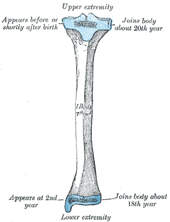
 Plan of ossification of the tibia. From three centers.
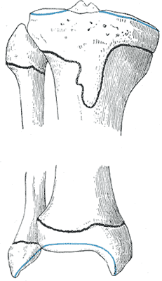
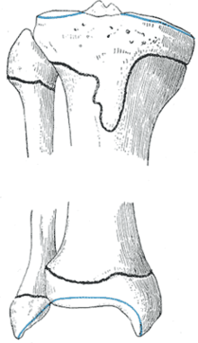 Epiphysial lines of tibia and fibula in a young adult. Anterior aspect.
The tibia (a bone in the Human Body) is ossified from three centers : one for the body and one for either extremity. Ossification begins in the center of the body, about the seventh week of fetal life, and gradually extends toward the extremities.
The center for the upper epiphysis appears before or shortly after birth at close to 34 weeks gestation; it is flattened in form, and has a thin tongue-shaped process in front, which forms the tuberosity; that for the lower epiphysis appears in the second year.
The lower epiphysis fuses with the tibial shaft at about the eighteenth, and the upper one fuses about the twentieth year.
Two additional centers occasionally exist, one for the tongue-shaped process of the upper epiphysis, which forms the tuberosity, and one for the medial malleolus.
This article was originally based on an entry from a public domain edition of Gray's Anatomy. As such, some of the information contained within it may be outdated.
Embryology of bones, joints, and muscles (GA 2.80, TE E5.0-2) Ossification Lower limbOssification of tibiaHeadcranium: Ossification of occipital bone · Ossification of frontal bone · Ossification of temporal bone · Ossification of sphenoid · Ossification of ethmoid
facial bones: Ossification of vomer (Sutura vomerina · Foramen vomerinum · Meatus vomerinus · Fissura vomerina) · Ossification of maxilla · Ossification of mandibleOtherOther M: JNT
anat(h/c, u, t, l)/phys
noco(arth/defr/back/soft)/cong, sysi/epon, injr
proc, drug(M01C, M4)
Categories:- Bones of the lower limb
Wikimedia Foundation. 2010.