- Ossification of humerus
-
 Figure 4 : Plan of ossification of the humerus.
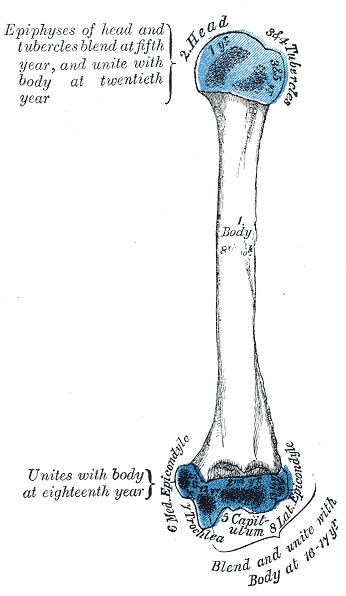
 Figure 5 : Epiphysial lines of humerus in a young adult. Anterior aspect. The lines of attachment of the articular capsules are in blue.
The humerus is ossified from eight centers, one for each of the following parts: the body, the head, the greater tubercle, the lesser tubercle, the capitulum, the trochlea, and one for each epicondyle. One primary and seven secondary centres .
The center for the body appears near the middle of the diaphysis in the eighth week of fetal life, and soon extends toward the extremities. This is the primary centre
At birth the humerus is ossified in nearly its whole length, only the extremities remaining cartilaginous.
During the first year, sometimes before birth, ossification commences in the head of the bone, and during the second year the center for the greater tubercle, and during the fifth that for the lesser tubercle, make their appearance.
By the sixth year the centers for the head and tubercles have joined, so as to form a single large epiphysis, which fuses with the body about the twentieth year.
The lower end of the humerus is ossified as follows.
At the end of the second year ossification begins in the capitulum, and extends medialward, to form the chief part of the articular end of the bone; the center for the medial part of the trochlea appears about the age of twelve.
Ossification begins in the medial epicondyle about the fifth year, and in the lateral about the thirteenth or fourteenth year.
About the sixteenth or seventeenth year, the lateral epicondyle and both portions of the articulating surface, having already joined, unite with the body, and at the eighteenth year the medial epicondyle becomes joined to it.
Embryology of bones, joints, and muscles (GA 2.80, TE E5.0-2) Ossification Lower limbHeadcranium: Ossification of occipital bone · Ossification of frontal bone · Ossification of temporal bone · Ossification of sphenoid · Ossification of ethmoid
facial bones: Ossification of vomer (Sutura vomerina · Foramen vomerinum · Meatus vomerinus · Fissura vomerina) · Ossification of maxilla · Ossification of mandibleOtherOther M: JNT
anat(h/c, u, t, l)/phys
noco(arth/defr/back/soft)/cong, sysi/epon, injr
proc, drug(M01C, M4)
Categories:- Skeletal disorders
Wikimedia Foundation. 2010.