Ossification of ethmoid

Ossification of ethmoid
Bone: Ossification of ethmoid
Gray194.png
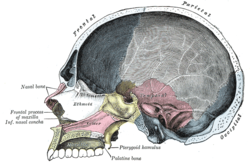
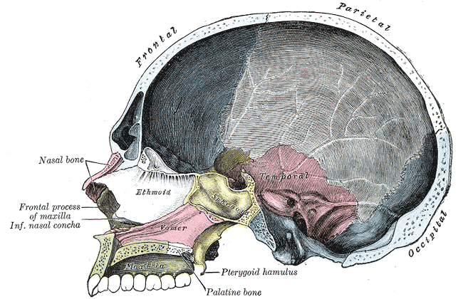
Sagittal section of skull. (Ethmoid bone visible as white structure to left.)
Gray's subject #36 153

The ethmoid is ossified in the cartilage of the nasal capsule by three centers: one for the perpendicular plate, and one for each labyrinth.

The labyrinths are first developed, ossific granules making their appearance in the region of the lamina papyracea between the fourth and fifth months of fetal life, and extending into the conchæ.

At birth, the bone consists of the two labyrinths, which are small and ill-developed. During the first year after birth, the perpendicular plate and crista galli begin to ossify from a single center, and are joined to the labyrinths about the beginning of the second year.

The cribriform plate is ossified partly from the perpendicular plate and partly from the labyrinths.

The development of the ethmoidal cells begins during fetal life.

This article was originally based on an entry from a public domain edition of Gray's Anatomy. As such, some of the information contained within it may be outdated.


Wikimedia Foundation. 2010.

Игры ⚽ Поможем сделать НИР

Look at other dictionaries:

  • Ossification center — Section of fetal bone of cat. ir. Irruption of the subperiosteal tissue. p. Fibrous layer of the periosteum. o. Layer of osteoblasts. im. Subperiosteal bony deposit. Latin centrum ossificationis Gray …   Wikipedia

  • Ossification of sphenoid — Figure 4 : Sphenoid bone at birth, posterior aspect. Until the seventh or eighth month of fetal life the body of the sphenoid consists of two parts: one in front of the tuberculum sellæ, the presphenoid, with which the small wings are… …   Wikipedia

  • Ossification of vomer — Vomer of infant. At an early period the septum of the nose consists of a plate of cartilage, the ethmovomerine cartilage. The postero superior part of this cartilage is ossified to form the perpendicular plate of the ethmoid; its antero inferior… …   Wikipedia

  • Ethmoid bone — Bone: Ethmoid bone Cranial bones …   Wikipedia

  • Ossification — Bone is broken down by osteoclasts, and rebuilt by osteoblasts, both of which communicate through cytokine (TGF β, IGF) signalling. Ossification (or osteogenesis) is the process of laying down new bone material by cells called osteoblasts. It is… …   Wikipedia

  • Ossification of scapula — Figure 5 : Plan of ossification of the scapula. From seven centers. The scapula is ossified from 7 or more centers: one for the body, two for the coracoid process, two for the acromion, one for the vertebral border, and one for the inferior… …   Wikipedia

  • Ossification of humerus — Figure 4 : Plan of ossification of the humerus. Figure 5 : Epiphysial lines of humerus in a young adult. Anterior aspect. The lines of attachment of the articular …   Wikipedia

  • Ossification of mandible — Figure 3: Mandible of human embryo 24 mm. long. Outer aspect. Figure 4: Mandible of human embryo 24 mm. long. Inner …   Wikipedia

  • Ossification of frontal bone — Figure 3: Frontal bone at birth The frontal bone is ossified in membrane from two primary centers, one for each half, which appear toward the end of the second month of fetal life, one above each supraorbital margin. From each of these centers,… …   Wikipedia

  • Ossification of tibia — Plan of ossification of the tibia. From three centers. Epiphysial lines of tibia and fibula in a young adult. Anter …   Wikipedia

Share the article and excerpts

Direct link
Do a right-click on the link above
and select “Copy Link”