- Ossification of temporal bone
-
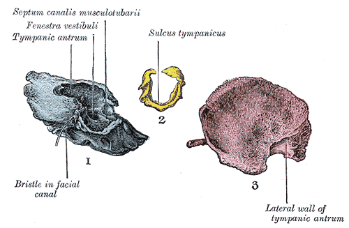
 1. Outer surface of petromastoid part. 2. Outer surface of tympanic ring. 3. Inner surface of squama.
 Figure 7 : Temporal bone at birth. Outer aspect.
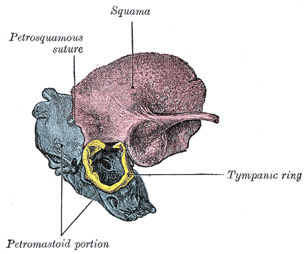
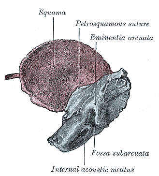
 Figure 8 : Temporal bone at birth. Inner aspect.
The temporal bone is ossified from eight centers, exclusive of those for the internal ear and the tympanic ossicles: one for the squama including the zygomatic process, one for the tympanic part, four for the petrous and mastoid parts, and two for the styloid process. Just before the close of fetal life [Fig. 6] the temporal bone consists of three principal parts:
- The squama is ossified in membrane from a single nucleus, which appears near the root of the zygomatic process about the second month.
- The petromastoid part is developed from four centers, which make their appearance in the cartilaginous ear capsule about the fifth or sixth month. One (proötic) appears in the neighborhood of the eminentia arcuata, spreads in front and above the internal acoustic meatus and extends to the apex of the bone; it forms part of the cochlea, vestibule, superior semicircular canal, and medial wall of the tympanic cavity. A second (opisthotic) appears at the promontory on the medial wall of the tympanic cavity and surrounds the fenestra cochleæ; it forms the floor of the tympanic cavity and vestibule, surrounds the carotid canal, invests the lateral and lower part of the cochlea, and spreads medially below the internal acoustic meatus. A third (pterotic) roofs in the tympanic cavity and antrum; while the fourth (epiotic) appears near the posterior semicircular canal and extends to form the mastoid process (Vrolik).
- The tympanic ring is an incomplete circle, in the concavity of which is a groove, the tympanic sulcus, for the attachment of the circumference of the tympanic membrane. This ring expands to form the tympanic part, and is ossified in membrane from a single center which appears about the third month. The styloid process is developed from the proximal part of the cartilage of the second branchial or hyoid arch by two centers: one for the proximal part, the tympanohyal, appears before birth; the other, comprising the rest of the process, is named the stylohyal, and does not appear until after birth. The tympanic ring unites with the squama shortly before birth; the petromastoid part and squama join during the first year, and the tympanohyal portion of the styloid process about the same time [Fig. 7, 8]. The stylohyal does not unite with the rest of the bone until after puberty, and in some skulls never at all.
The chief subsequent changes in the temporal bone apart from increase in size are:
- The tympanic ring extends outward and backward to form the tympanic part. This extension does not, however, take place at an equal rate all around the circumference of the ring, but occurs most rapidly on its anterior and posterior portions, and these outgrowths meet and blend, and thus, for a time, there exists in the floor of the meatus a foramen, the foramen of Huschke; this foramen is usually closed about the fifth year, but may persist throughout life.
- The mandibular fossa is at first extremely shallow, and looks lateralward as well as downward; it becomes deeper and is ultimately directed downward. Its change in direction is accounted for as follows. The part of the squama which forms the fossa lies at first below the level of the zygomatic process. As, however, the base of the skull increases in width, this lower part of the squama is directed horizontally inward to contribute to the middle fossa of the skull, and its surfaces therefore come to look upward and downward; the attached portion of the zygomatic process also becomes everted, and projects like a shelf at right angles to the squama.
- The mastoid portion is at first quite flat, and the stylomastoid foramen and rudimentary styloid process lie immediately behind the tympanic ring. With the development of the air cells the outer part of the mastoid portion grows downward and forward to form the mastoid process, and the styloid process and stylomastoid foramen now come to lie on the under surface. The descent of the foramen is necessarily accompanied by a corresponding lengthening of the facial canal.
- The downward and forward growth of the mastoid process also pushes forward the tympanic part, so that the portion of it which formed the original floor of the meatus and contained the foramen of Huschke is ultimately found in the anterior wall.
- The fossa subarcuata becomes filled up and almost obliterated.
Embryology of bones, joints, and muscles (GA 2.80, TE E5.0-2) Ossification Lower limbHeadcranium: Ossification of occipital bone · Ossification of frontal bone · Ossification of temporal bone · Ossification of sphenoid · Ossification of ethmoid
facial bones: Ossification of vomer (Sutura vomerina · Foramen vomerinum · Meatus vomerinus · Fissura vomerina) · Ossification of maxilla · Ossification of mandibleOtherOther M: JNT
anat(h/c, u, t, l)/phys
noco(arth/defr/back/soft)/cong, sysi/epon, injr
proc, drug(M01C, M4)
Categories:- Skeletal disorders
Wikimedia Foundation. 2010.