- Ossification of occipital bone
-
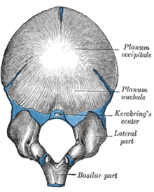 Figure 3 : Occipital bone at birth.
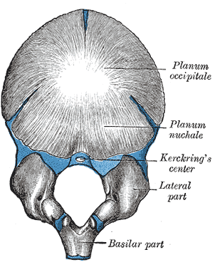
The planum occipitale [Fig. 3] of the squama of the occipital bone is developed in membrane, and may remain separate throughout life when it constitutes the interparietal bone; the rest of the bone is developed in cartilage.
The number of nuclei for the planum occipitale is usually given as four, two appearing near the middle line about the second month, and two some little distance from the middle line about the third month of fetal life.
The planum nuchale of the squama is ossified from two centers, which appear about the seventh week of fetal life and soon unite to form a single piece.
Union of the upper and lower portions of the squama takes place in the third month of fetal life.
An occasional center (Kerckring) appears in the posterior margin of the foramen magnum during the fifth month; this forms a separate ossicle (sometimes double) which unites with the rest of the squama before birth.
Each of the lateral parts begins to ossify from a single center during the eighth week of fetal life. The basilar portion is ossified from two centers, one in front of the other; these appear about the sixth week of fetal life and rapidly coalesce.
Mall states that the planum occipitale is ossified from two centers and the basilar portion from one.
About the fourth year the squama and the two lateral portions unite, and about the sixth year the bone consists of a single piece. Between the 18th and 25th years the occipital and sphenoid become united, forming a single bone.
In other animals
In many animals these parts stay separate through life; e.g. in the dog as four parts: supraoccipital (= the squama); left and right exoccipital (= the lateral parts); basioccipital (= the basilar part).
This article was originally based on an entry from a public domain edition of Gray's Anatomy. As such, some of the information contained within it may be outdated.
Embryology of bones, joints, and muscles (GA 2.80, TE E5.0-2) Ossification Lower limbHeadcranium: Ossification of occipital bone · Ossification of frontal bone · Ossification of temporal bone · Ossification of sphenoid · Ossification of ethmoid
facial bones: Ossification of vomer (Sutura vomerina · Foramen vomerinum · Meatus vomerinus · Fissura vomerina) · Ossification of maxilla · Ossification of mandibleOtherOther M: JNT
anat(h/c, u, t, l)/phys
noco(arth/defr/back/soft)/cong, sysi/epon, injr
proc, drug(M01C, M4)
Categories:- Skeletal system
- Skull
Wikimedia Foundation. 2010.