- Ossification of scapula
-
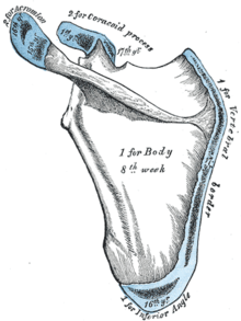 Figure 5 : Plan of ossification of the scapula. From seven centers.
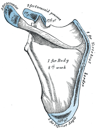
The scapula is ossified from 7 or more centers: one for the body, two for the coracoid process, two for the acromion, one for the vertebral border, and one for the inferior angle.
Ossification of the body begins about the second month of fetal life, by an irregular quadrilateral plate of bone forming, immediately behind the glenoid cavity. This plate extends to form the chief part of the bone, the scapular spine growing up from its dorsal surface about the third month. Ossification starts as membranous ossification before birth[1][2]. After birth, the cartilaginous components would undergo endochondral ossification.
At birth, a large part of the scapula is osseous, but the glenoid cavity, the coracoid process, the acromion, the vertebral border, and the inferior angle are cartilaginous.
From the 15th to the 18th month after birth, ossification takes place in the middle of the coracoid process, which as a rule becomes joined with the rest of the bone about the 15th year.
Between the 14th and 20th months, the remaining parts ossify in quick succession, and usually in this order; first, in the root of the coracoid process, in the form of a broad scale; secondly, near the base of the acromion; thirdly, in the inferior angle and contiguous part of the vertebral border; fourthly, near the extremity of the acromion; fifthly, in the vertebral border.
The base of the acromion is formed by an extension from the spine; the two nuclei of the acromion unite, and then join with the extension from the spine. The upper third of the glenoid cavity is ossified from a separate center (sub coracoid), which appears between the 10th and 11th years and joins between the 16th and the 18th years.
Further, an epiphysial plate appears for the lower part of the glenoid cavity, and the tip of the coracoid process frequently has a separate nucleus. These various epiphyses are joined to the bone by the 25th year.
Failure of bony union between the acromion and spine sometimes occurs (see os acromiale), the junction being effected by fibrous tissue, or by an imperfect articulation; in some cases of supposed fracture of the acromion with ligamentous union, it is probable that the detached segment was never united to the rest of the bone.
References
Embryology of bones, joints, and muscles (GA 2.80, TE E5.0-2) Ossification Lower limbHeadcranium: Ossification of occipital bone · Ossification of frontal bone · Ossification of temporal bone · Ossification of sphenoid · Ossification of ethmoid
facial bones: Ossification of vomer (Sutura vomerina · Foramen vomerinum · Meatus vomerinus · Fissura vomerina) · Ossification of maxilla · Ossification of mandibleOtherOssification of scapulaOther M: JNT
anat(h/c, u, t, l)/phys
noco(arth/defr/back/soft)/cong, sysi/epon, injr
proc, drug(M01C, M4)
Categories:- Skeletal disorders
Wikimedia Foundation. 2010.