- Coracoid process
-
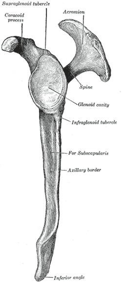
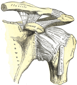
Bone: coracoid process Left scapula. Lateral view. (Coracoid process labeled at upper left.) The left shoulder and acromioclavicular joints, and the proper ligaments of the scapula. (Coracoid process visible at center.) Anterior view. Latin processus coracoideus Gray's subject #50 207 The coracoid process (from greek κόραξ, crow) is a small hook-like structure on the lateral edge of the superior anterior portion of the scapula. Pointing laterally forward, it, together with the acromion, serves to stabilize the shoulder joint. It is palpable in the deltopectoral groove between the deltoid and pectoralis major muscles.
"Coracoid" in itself means "like a raven's beak", with reference to its shape. (Greek "Korax" = Raven)
Contents
Human anatomy
Attachments
It is the site of attachment for several structures:
- The pectoralis minor muscle - to 3rd to 5th ribs.
- The short head of biceps brachii muscle - to Radial tuberosity.
- The coracobrachialis muscle - to medial humerus.
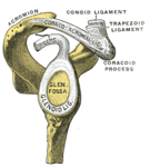
- The coracoclavicular ligament - to the clavicle. (The ligament is formed by the conoid ligament and trapezoid ligament.)
- The coracoacromial ligament - to the acromion
- The coracohumeral ligament - to the humerus
- The superior transverse scapular ligament - from the base of the coracoid to the medial portion of the suprascapular notch
Structure
The coracoid process is a thick curved process attached by a broad base to the upper part of the neck of the scapula; it runs at first upward and medialward; then, becoming smaller, it changes its direction, and projects forward and lateralward.
The ascending portion, flattened from before backward, presents in front a smooth concave surface, across which the Subscapularis passes.
The horizontal portion is flattened from above downward; its upper surface is convex and irregular, and gives attachment to the Pectoralis minor; its under surface is smooth; its medial and lateral borders are rough; the former gives attachment to the Pectoralis minor and the latter to the coracoacromial ligament; the apex is embraced by the conjoined tendon of origin of the Coracobrachialis and short head of the Biceps brachii and gives attachment to the coracoclavicular fascia.
On the medial part of the root of the coracoid process is a rough impression for the attachment of the conoid ligament; and running from it obliquely forward and lateralward, on to the upper surface of the horizontal portion, is an elevated ridge for the attachment of the trapezoid ligament.
Clinical relevance
The coracoid process is palpable just below the lateral end of the clavicle (collar bone). It is otherwise known as the "Surgeon's Lighthouse" because it serves as a landmark to avoid neurovascular damage.[2] Major neurovascular structures enter the upper limb medial to the coracoid process so surgical approaches to the shoulder region always takes place lateral to the coracoid process.
Evolutionary variation
In monotremes, the coracoid is a separate bone. Reptiles, birds, and frogs (but not salamanders) also possess a bone by this name, but is not homologous with the coracoid process of mammals.[3]
Analyses of the size and shape of the coracoid process in Australopithecus africanus (STS 7) have shown that in this species it displayed a prominent dorsolateral tubercle placed more laterally than in modern humans. This reflect, according to one interpretation, a scapula positioned high on a funnel-shaped thorax and a clavicle positioned obliquely as in extant great apes. [4]
Additional images
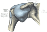
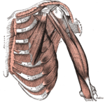
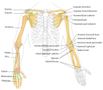
Capsule of shoulder-joint (distended). Anterior aspect.Glenoid fossa of right side.Deep muscles of the chest and front of the arm, with the boundaries of the axilla.Human arm bones diagramReferences
- ^ Gray's Anatomy (1918), see infobox.
- ^ Gallino, Mario; Santamaria, Eliana; Tiziana, Doro (1998). "Anthropometry of the scapula: Clinical and surgical considerations". Journal of Shoulder and Elbow Surgery 7 (3): 284–291. doi:10.1016/S1058-2746(98)90057-X. ""...defined by Matsen et al. as 'the lighthouse of the shoulder.'"
-
- Matsen, Frederick A; Lippitt, Steven B; Sidles, John A; Douglas T, Harryman (1984). Practical Evaluation and Management of the Shoulder. ISBN 978-0721648194.
-
- ^ Romer, Alfred Sherwood; Parsons, Thomas S. (1977). The Vertebrate Body. Philadelphia, PA: Holt-Saunders International. pp. 186–187. ISBN 0-03-910284-X.
- ^ Larson, Susan G. (2009). "Evolution of the Hominin Shoulder: Early Homo". In Grine, Frederick E.; Fleagle, John G.; Leakey, Richard E.. The First Humans - Origin and Early Evolution of the Genus Homo. Springer. pp. 65–6. doi:10.1007/978-1-4020-9980-9. ISBN 978-1-4020-9979-3. http://www.springerlink.com/content/p22t46/#section=62942&page=1.
External links
- coracoid+process at eMedicine Dictionary
- Bioweb at UWLAX aplab
- Anatomy at PSU skel/scapula2
Bones of upper limbs (TA A02.4, GA 2.200–230) Pectoral girdle,
clavicleScapula fossae (subscapular, supraspinatous, infraspinatous) · scapular notch · glenoid cavity
tubercles (infraglenoid, supraglenoid) · spine of scapula · acromion · coracoid process
borders (superior, lateral/axillary, medial/vertebral) · angles (superior, inferior, lateral)Humerus upper extremity: necks (anatomical, surgical) · tubercles (greater, lesser) · intertubercular sulcus
body: radial sulcus · deltoid tuberosity
lower extremity: capitulum · trochlea · epicondyles (lateral, medial) · supracondylar ridges (lateral, medial) · fossae (radial, coronoid, olecranon)Forearm Hand carpus: scaphoid · lunate · triquetral · pisiform · trapezium · trapezoid · capitate · hamate (hamulus)
metacarpus: 1st metacarpal · 2nd · 3rd · 4th · 5th
phalanges of the hand: proximal · intermediate · distalCategories:- Bones of the upper limb
- Musculoskeletal system stubs
Wikimedia Foundation. 2010.

