- Body of radius
Infobox Bone
Name = Body of radius
Latin = corpus radii
GraySubject = 52
GrayPage = 219
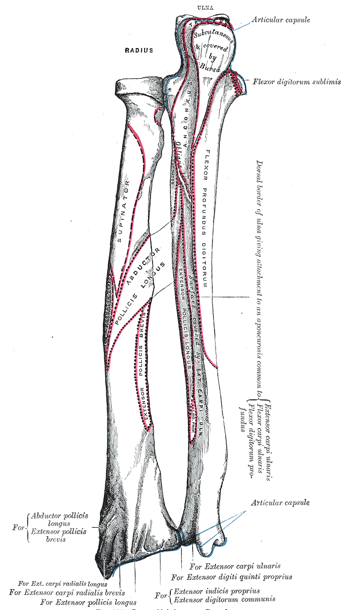
Caption = The "radius" andulna of the left forearm, posterior surface. The top is proximal (elbow) and bottom is distal (wrist).
Caption2 =
Precursor =
System =
Artery =
Vein =
Nerve =
Lymph =
MeshName =
MeshNumber =
DorlandsPre = c_56
DorlandsSuf = 12260789
The body of the radius (or shaft of radius) is prismoid in form, narrower above than below, and slightly curved, so as to be convex lateralward. It presents three borders and three surfaces.Borders
The volar border ("margo volaris; anterior border") extends from the lower part of the tuberosity above to the anterior part of the base of the styloid process below, and separates the volar from the lateral surface. Its upper third is prominent, and from its oblique direction has received the name of the oblique line of the radius; it gives origin to the
flexor digitorum superficialis muscle (also "flexor digitorum sublimis") andflexor pollicis longus muscle ; the surface above the line gives insertion to part of thesupinator muscle . The middle third of the volar border is indistinct and rounded. The lower fourth is prominent, and gives insertion to thepronator quadratus muscle , and attachment to the dorsal carpal ligament; it ends in a small tubercle, into which the tendon of thebrachioradialis muscle is inserted.The dorsal border ("margo dorsalis; posterior border") begins above at the back of the neck, and ends below at the posterior part of the base of the styloid process; it separates the posterior from the lateral surface. is indistinct above and below, but well-marked in the middle third of the bone.
The interosseous crest ("crista interossea; internal or interosseous border") begins above, at the back part of the tuberosity, and its upper part is rounded and indistinct; it becomes sharp and prominent as it descends, and at its lower part divides into two ridges which are continued to the anterior and posterior margins of the ulnar notch. To the posterior of the two ridges the lower part of the
interosseous membrane is attached, while the triangular surface between the ridges gives insertion to part of thepronator quadratus muscle . This crest separates the volar from the dorsal surface, and gives attachment to the interosseous membrane. The connection between the two bones is actually a joint referred to as asyndesmoses joint.Surface
The volar surface ("facies volaris; anterior surface") is concave in its upper three-fourths, and gives origin to the
flexor pollicis longus muscle ; it is broad and flat in its lower fourth, and affords insertion to the Pronator quadratus. A prominent ridge limits the insertion of the Pronator quadratus below, and between this and the inferior border is a triangular rough surface for the attachment of the volar radiocarpal ligament. At the junction of the upper and middle thirds of the volar surface is the nutrient foramen, which is directed obliquely upward.The dorsal surface ("facies dorsalis; posterior surface") is convex, and smooth in the upper third of its extent, and covered by the Supinator. Its middle third is broad, slightly concave, and gives origin to the Abductor pollicis longus above, and the
extensor pollicis brevis muscle below. Its lower third is broad, convex, and covered by the tendons of the muscles which subsequently run in the grooves on the lower end of the bone.The lateral surface ("facies lateralis; external surface") is convex throughout its entire extent. Its upper third gives insertion to the supinator muscle. About its center is a rough ridge, for the insertion of the
pronator teres muscle . Its lower part is narrow, and covered by the tendons of theabductor pollicis longus muscle andextensor pollicis brevis muscle .
Wikimedia Foundation. 2010.