- Deltoid tuberosity
-
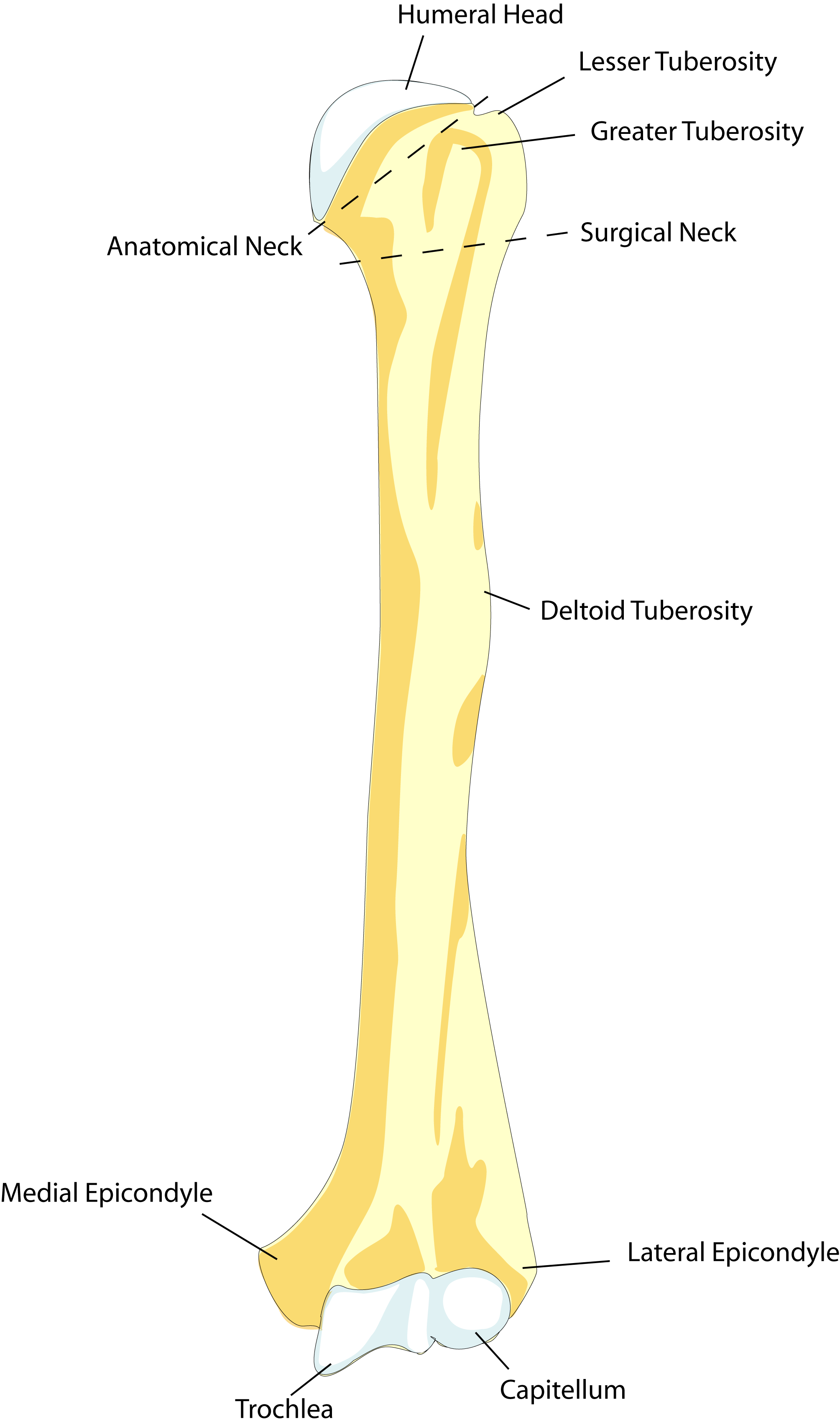
Bone: Deltoid tuberosity 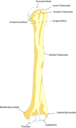
Left humerus. Anterior view. (Deltoideus labeled at center right.) Latin tuberositas deltoidea humeri Gray's subject #51 211 In human anatomy, the deltoid tuberosity is a rough, triangular[1] area on the anterolateral (exterior-front) surface of the middle of the humerus to which the deltoid muscle attaches. [2]
Contents
Variation
It has been reported as very prominent in less than 10% of cases. [3]
Development
The deltoid tuberosity develops through endochondral ossification in a two-phase process. The initiating signal is tendon-dependent, whilst the growth phase is muscle-dependent. [4]
Evolutionary variation
In mammals, the humerus displays a wide morphological variation. The size and orientation of its functionally important features, including the deltoid tubercle, greater tubercle, and medial epicondyle, are pivotal to an animal's style of locomotion and habitat. In cursorial (running) animals such as the Pronghorn Antelope, the deltoid tubercle is located about a quarter of the way down the shaft, which allows for rapid but relatively weak limb flexion and extension. In natatorial (swimming) animals such as the North American River Otter, the tubercle is located nearly half way down the shaft, which allows for powerful limb flexion and extension. The tuberosity can be very pronounced in fossorial (digging) animals, such as the Mountain Beaver. [5]
See also
- Deltoid tubercle (on clavicle)
References
- ^ Gray, Henry (1918). Gray's Anatomy. ISBN 1859580181. (See link in infobox.)
- ^ Feneis, Heinz (2000). Pocket Atlas of Human Anatomy (4th ed.). Thieme. p. 36. ISBN 3-13-511204-7.
- ^ Fink-Bennett D, Vicuna-Rios J. (1980). "The deltoid tuberosity--a potential pitfall (the "delta sign") in bone-scan interpretation: concise communication". The Journal of Nuclear Medicine 21 (3): 211–212. http://jnm.snmjournals.org/cgi/reprint/21/3/211. "...in seven out of 100 scans reviewed."
- ^ Blitz E et al. (December 2009). "Bone ridge patterning during musculoskeletal assembly is mediated through SCX regulation of Bmp4 at the tendon-skeleton junction". Dev Cell 6 (17): 861–73. doi:10.1016/j.devcel.2009.10.010. PMID 20059955.
- ^ Hall, Brian Keith (2007). Fins into limbs: evolution, development, and transformation. University of Chicago Press. p. 251. ISBN 0226313379. http://books.google.com/books?id=Z0YWn5F9sWkC&pg=PA251. (Including an illustration of variation in mammalian humeri.)
Bones of upper limbs (TA A02.4, GA 2.200–230) Pectoral girdle,
clavicleScapula fossae (subscapular, supraspinatous, infraspinatous) · scapular notch · glenoid cavity
tubercles (infraglenoid, supraglenoid) · spine of scapula · acromion · coracoid process
borders (superior, lateral/axillary, medial/vertebral) · angles (superior, inferior, lateral)Humerus upper extremity: necks (anatomical, surgical) · tubercles (greater, lesser) · intertubercular sulcus
body: radial sulcus · deltoid tuberosity
lower extremity: capitulum · trochlea · epicondyles (lateral, medial) · supracondylar ridges (lateral, medial) · fossae (radial, coronoid, olecranon)Forearm Hand carpus: scaphoid · lunate · triquetral · pisiform · trapezium · trapezoid · capitate · hamate (hamulus)
metacarpus: 1st metacarpal · 2nd · 3rd · 4th · 5th
phalanges of the hand: proximal · intermediate · distalCategories:- Bones of the upper limb
Wikimedia Foundation. 2010.