Ossification of radius

Ossification of radius
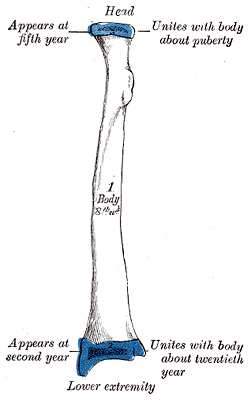
Figure 1 : Plan of ossification of the radius. From three centers.
Figure 2 : Epiphysial lines of radius in a young adult. Anterior aspect. The line of attachment of the articular capsule of the wrist-joint is in blue.

The radius is one of the two bones in the forearm.

The radius is ossified from three centers: one for the body, and one for either extremity. That for the body makes its appearance near the center of the bone, during the eighth week of fetal life.

About the end of year, ossification commences in the lower end; and at the fifth year, in the upper end.

The upper epiphysis fuses with the body at the age of seventeen or eighteen years, the lower about the age of twenty.

An additional center sometimes found in the radial tuberosity, appears about the fourteenth or fifteenth year.


Wikimedia Foundation. 2010.

Игры ⚽ Нужен реферат?

Look at other dictionaries:

  • Ossification center — Section of fetal bone of cat. ir. Irruption of the subperiosteal tissue. p. Fibrous layer of the periosteum. o. Layer of osteoblasts. im. Subperiosteal bony deposit. Latin centrum ossificationis Gray …   Wikipedia

  • Ossification — Bone is broken down by osteoclasts, and rebuilt by osteoblasts, both of which communicate through cytokine (TGF β, IGF) signalling. Ossification (or osteogenesis) is the process of laying down new bone material by cells called osteoblasts. It is… …   Wikipedia

  • Ossification of scapula — Figure 5 : Plan of ossification of the scapula. From seven centers. The scapula is ossified from 7 or more centers: one for the body, two for the coracoid process, two for the acromion, one for the vertebral border, and one for the inferior… …   Wikipedia

  • Ossification of humerus — Figure 4 : Plan of ossification of the humerus. Figure 5 : Epiphysial lines of humerus in a young adult. Anterior aspect. The lines of attachment of the articular …   Wikipedia

  • Ossification of mandible — Figure 3: Mandible of human embryo 24 mm. long. Outer aspect. Figure 4: Mandible of human embryo 24 mm. long. Inner …   Wikipedia

  • Ossification of ethmoid — Bone: Ossification of ethmoid Sagittal section of skull. (Ethmoid bone visible as white structure to left.) Gray s subject …   Wikipedia

  • Ossification of frontal bone — Figure 3: Frontal bone at birth The frontal bone is ossified in membrane from two primary centers, one for each half, which appear toward the end of the second month of fetal life, one above each supraorbital margin. From each of these centers,… …   Wikipedia

  • Ossification of tibia — Plan of ossification of the tibia. From three centers. Epiphysial lines of tibia and fibula in a young adult. Anter …   Wikipedia

  • Ossification of sphenoid — Figure 4 : Sphenoid bone at birth, posterior aspect. Until the seventh or eighth month of fetal life the body of the sphenoid consists of two parts: one in front of the tuberculum sellæ, the presphenoid, with which the small wings are… …   Wikipedia

  • Ossification of ulna — Plan of ossification of the ulna. From three centers. Epiphysial lines of ulna in a young adult. Lateral aspect. The lines of attachment of the articular capsules are in blue …   Wikipedia

Share the article and excerpts

Direct link
Do a right-click on the link above
and select “Copy Link”