- Paraxial mesoderm
Infobox Embryology
Name = PAGENAME
Latin =
GraySubject = 6
GrayPage = 50

Caption = Transverse section of a chick embryo of forty-five hours’ incubation.
* "Chordamesoderm :" yellow, atnotochord .
* "Paraxial mesoderm:" red, atsomite .
* "Intermediate mesoderm :" purple, nearWolffian duct .
* "Lateral plate mesoderm :" purple, near "Somatic mesoderm" and "Splanchic mesoderm".
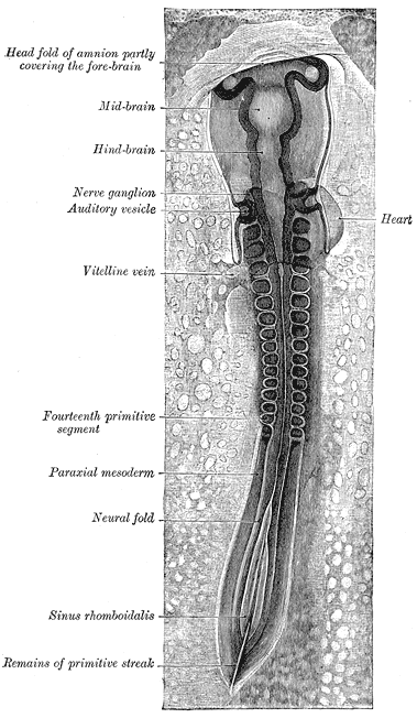
Caption2 = Chick embryo of thirty-three hours’ incubation, viewed from the dorsal aspect. (Paraxial mesoderm labeled at left.)
System =
CarnegieStage = 9
Precursor =
GivesRiseTo =somitomere ,head mesoderm
MeshName =
MeshNumber =
DorlandsPre = m_11
DorlandsSuf = 12527037Paraxial mesoderm is the area of
mesoderm that forms just lateral to theneural tube on both sides.It gives rise to the
somitomere s/somite s and mesoderm of thebranchial arches .*Somites form the
vertebral column ,dermis andskeletal muscle
*Branchial arches will develop intofacial muscle and cartilage, plus other structures.* eventually differentiates into the axial skeleton, skeletal muscle, part of the dermis
* almost immediately as it is formed, somitomeres develop.
* starts with several pairs in the cranial region, and increasingly more proceed to develop towardsthe caudal region.
* The original seven pairs form the straited muscles of head and neck, which develop within thepharyngeal arches
* The other somitomeres develop further, to form discrete blocks called somites, starting atapproximately 20 days.External links
* [http://www.ncbi.nlm.nih.gov/books/bv.fcgi?db=Books&rid=dbio.section.3455 Overview at nih.gov]
* [http://sprojects.mmi.mcgill.ca/embryology/earlydev/week4/somites.html Diagram and overview at mcgill.ca]
* [http://connection.lww.com/Products/sadler/images/figurelarge5-9.jpgDiagram at lww.com]
Wikimedia Foundation. 2010.