- Circulatory system
-
This article is about the organ system. For the band, see Circulatory System. For transport in plants, see Vascular tissue.
Circulatory system 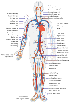
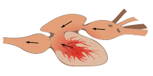
The human circulatory system. Red indicates oxygenated blood, blue indicates deoxygenated. Latin systema cardiovasculare The circulatory system is an organ system that passes nutrients (such as amino acids, electrolytes and lymph), gases, hormones, blood cells, etc. to and from cells in the body to help fight diseases and help stabilize body temperature and pH to maintain homeostasis.
This system may be seen strictly as a blood distribution network, but some consider the circulatory system as composed of the cardiovascular system, which distributes blood,[1] and the lymphatic system,[2] which distributes lymph. While humans, as well as other vertebrates, have a closed cardiovascular system (meaning that the blood never leaves the network of arteries, veins and capillaries), some invertebrate groups have an open cardiovascular system. The most primitive animal phyla lack circulatory systems. The lymphatic system, on the other hand, is an open system.
Two types of fluids move through the circulatory system: blood and lymph. The blood, heart, and blood vessels form the cardiovascular system. The lymph, lymph nodes, and lymph vessels form the lymphatic system. The cardiovascular system and the lymphatic system collectively make up the circulatory system.
Contents
Human cardiovascular system
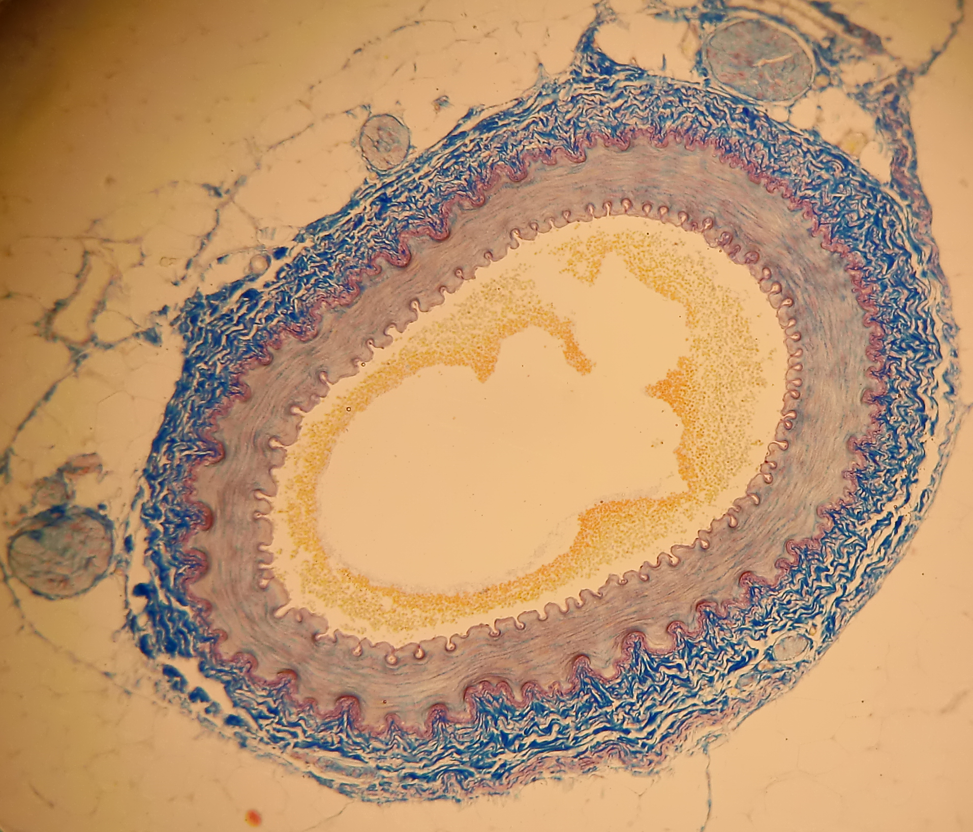
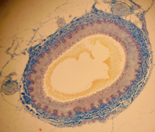 Cross section of a human artery
The main components of the human cardiovascular system are the heart, blood, and blood vessels.[3] It includes: the pulmonary circulation, a "loop" through the lungs where blood is oxygenated; and the systemic circulation, a "loop" through the rest of the body to provide oxygenated blood. An average adult contains five to six quarts (roughly 4.7 to 5.7 liters) of blood, which consists of plasma, red blood cells, white blood cells, and platelets. Also, the digestive system works with the circulatory system to provide the nutrients the system needs to keep the heart pumping.
Pulmonary circulation
Main article: Pulmonary circulationThe pulmonary circulatory system is the portion of the cardiovascular system in which oxygen-depleted blood is pumped away from the heart, via the pulmonary artery, to the lungs and returned, oxygenated, to the heart via the pulmonary vein.
Oxygen deprived blood from the vena cava, enters the right atrium of the heart and flows through the tricuspid valve (right atrioventricular valve) into the right ventricle, from which it is then pumped through the pulmonary semilunar valve into the pulmonary artery to the lungs. Gas exchange occurs in the lungs, whereby CO2 is released from the blood, and oxygen is absorbed. The pulmonary vein returns the now oxygen-rich blood to the heart.
Systemic circulation
Main article: Systemic circulationSystemic circulation is the portion of the cardiovascular system which transports oxygenated blood away from the heart, to the rest of the body, and returns oxygen-depleted blood back to the heart. Systemic circulation is, distance-wise, much longer than pulmonary circulation, transporting blood to every part of the body.
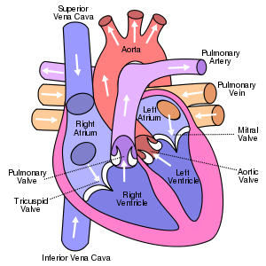 View from the front, which means the right side of the heart is on the left of the diagram (and vice-versa)
Coronary circulation
Main article: Coronary circulationThe coronary circulatory system provides a blood supply to the heart. As it provides oxygenated blood to the heart, it is by definition a part of the systemic circulatory system.
Heart
Main article: HeartThe heart pumps oxygenated blood to the body and deoxygenated blood to the lungs. In the human heart there is one atrium and one ventricle for each circulation, and with both a systemic and a pulmonary circulation there are four chambers in total: left atrium, left ventricle, right atrium and right ventricle. The right atrium is the upper chamber of the right side of the heart. The blood that is returned to the right atrium is deoxygenated (poor in oxygen) and passed into the right ventricle to be pumped through the pulmonary artery to the lungs for re-oxygenation and removal of carbon dioxide. The left atrium receives newly oxygenated blood from the lungs as well as the pulmonary vein which is passed into the strong left ventricle to be pumped through the aorta to the different organs of the body.
Closed cardiovascular system
The cardiovascular systems of humans are closed, meaning that the blood never leaves the network of blood vessels. In contrast, oxygen and nutrients diffuse across the blood vessel layers and enters interstitial fluid, which carries oxygen and nutrients to the target cells, and carbon dioxide and wastes in the opposite direction. The other component of the circulatory system, the lymphatic system, is not closed.
Oxygen transportation
Main article: Blood#Oxygen transportAbout 98.5% of the oxygen in a sample of arterial blood in a healthy human breathing air at sea-level pressure is chemically combined with haemoglobin molecules. About 1.5% is physically dissolved in the other blood liquids and not connected to haemoglobin. The haemoglobin molecule is the primary transporter of oxygen in mammals and many other species.
 An animation of a typical human red blood cell cycle in the circulatory system. This animation occurs at real time (20 seconds of cycle) and shows the red blood cell deform as it enters capillaries, as well as changing color as it alternates in states of oxygenation along the circulatory system.
Development
The development of the circulatory system initially occurs by the process of vasculogenesis. The human arterial and venous systems develop from different embryonic areas. While the arterial system develops mainly from the aortic arches, the venous system arises from three bilateral veins during weeks 4 - 8 of human development.
Arterial development
Main article: Aortic archesThe human arterial system originate from the aortic arches and from the dorsal aortae starting from week 4 of human development. Aortic arch 1 almost completely regresses except to form the maxillary arteries. Aortic arch 2 also completely regresses except to form the stapedial arteries. The definitive formation of the arterial system arise from aortic arches 3, 4 and 6. While aortic arch 5 completely regreses.
The dorsal aortae are initially bilateral and then fuse to form the definitive dorsal aorta. Approximately 30 posterolateral branches arise off the aorta and will form the intercostal arteries, upper and lower extremity arteries, lumbar arteries and the lateral sacral arteries. The lateral branches of the aorta form the definitive renal, suprarrenal and gonadal arteries. Finally, the ventral branches of the aorta consist of the vitelline arteries and umbilical arteries. The vitelline arteries form the celiac, superior and inferior mesenteric arteries of the gastrointestinal tract. After birth, the umbilical arteries will form the internal iliac arteries.
Venous development
The human venous system develops mainly from the vitelline veins, the umbilical veins and the cardinal veins, all of which empty into the sinus venosus.
Measurement techniques
- Electrocardiogram—for cardiac electrophysiology
- Sphygmomanometer and stethoscope—for blood pressure
- Pulse meter—for cardiac function (heart rate, rhythm, dropped beats)
- Pulse—commonly used to determine the heart rate in absence of certain cardiac pathologies
- Heart rate variability -- used to measure variations of time intervals between heart beats
- Nail bed blanching test—test for perfusion
- Vessel cannula or catheter pressure measurement—pulmonary wedge pressure or in older animal experiments.
Health and disease
Main article: Cardiovascular diseaseMain article: Congenital heart defectNonhuman
Other vertebrates
 Two-chambered heart of a fish
The circulatory systems of all vertebrates, as well as of annelids (for example, earthworms) and cephalopods (squid and octopus) are closed, just as in humans. Still, the systems of fish, amphibians, reptiles, and birds show various stages of the evolution of the circulatory system.
In fish, the system has only one circuit, with the blood being pumped through the capillaries of the gills and on to the capillaries of the body tissues. This is known as single cycle circulation. The heart of fish is therefore only a single pump (consisting of two chambers).
In amphibians and most reptiles, a double circulatory system is used, but the heart is not always completely separated into two pumps. Amphibians have a three-chambered heart.
In reptiles, the ventricular septum of the heart is incomplete and the pulmonary artery is equipped with a sphincter muscle. This allows a second possible route of blood flow. Instead of blood flowing through the pulmonary artery to the lungs, the sphincter may be contracted to divert this blood flow through the incomplete ventricular septum into the left ventricle and out through the aorta. This means the blood flows from the capillaries to the heart and back to the capillaries instead of to the lungs. This process is useful to ectothermic (cold-blooded) animals in the regulation of their body temperature.
Birds and mammals show complete separation of the heart into two pumps, for a total of four heart chambers; it is thought[citation needed] that the four-chambered heart of birds evolved independently from that of mammals.
Open circulatory system
The open circulatory system is a system in which fluid (called hemolymph) in a cavity called the hemocoel bathes the organs directly with oxygen and nutrients and there is no distinction between blood and interstitial fluid; this combined fluid is called hemolymph or haemolymph. Muscular movements by the animal during locomotion can facilitate hemolymph movement, but diverting flow from one area to another is limited. When the heart relaxes, blood is drawn back toward the heart through open-ended pores (ostia).
Hemolymph fills all of the interior hemocoel of the body and surrounds all cells. Hemolymph is composed of water, inorganic salts (mostly Na+, Cl-, K+, Mg2+, and Ca2+), and organic compounds (mostly carbohydrates, proteins, and lipids). The primary oxygen transporter molecule is hemocyanin.
There are free-floating cells, the hemocytes, within the hemolymph. They play a role in the arthropod immune system.
Absence of circulatory system
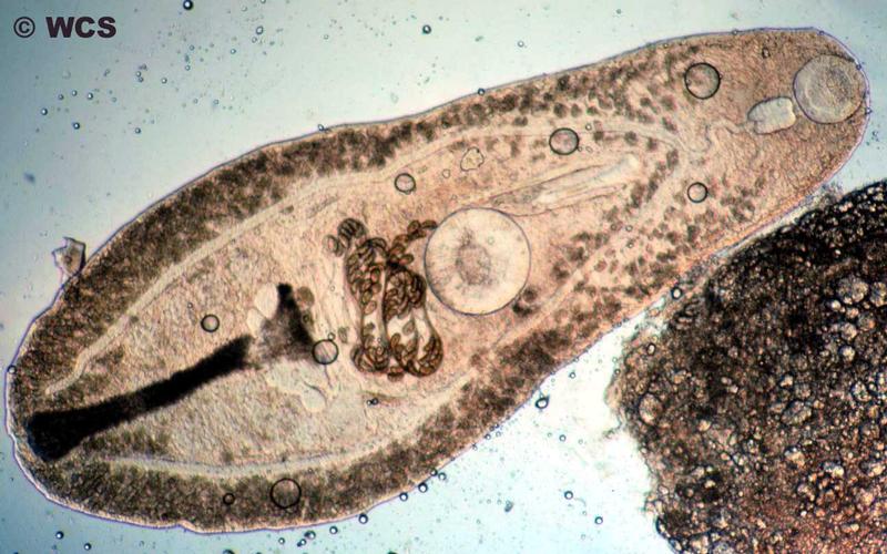
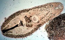 Flatworms, such as this Helicometra sp., lack specialized circulatory organs
Circulatory systems are absent in some animals, including flatworms (phylum Platyhelminthes). Their body cavity has no lining or enclosed fluid. Instead a muscular pharynx leads to an extensively branched digestive system that facilitates direct diffusion of nutrients to all cells. The flatworm's dorso-ventrally flattened body shape also restricts the distance of any cell from the digestive system or the exterior of the organism. Oxygen can diffuse from the surrounding water into the cells, and carbon dioxide can diffuse out. Consequently every cell is able to obtain nutrients, water and oxygen without the need of a transport system.
Some animals, such as jellyfish, have more extensive branching from their gastrovascular cavity (which functions as both a place of digestion and a form of circulation), this branching allows for bodily fluids to reach the outer layers, since the digestion begins in the inner layers.
History of discovery
The earliest known writings on the circulatory system are found in the Ebers Papyrus (16th century BCE), an ancient Egyptian medical papyrus containing over 700 prescriptions and remedies, both physical and spiritual. In the papyrus, it acknowledges the connection of the heart to the arteries. The Egyptians thought air came in through the mouth and into the lungs and heart. From the heart, the air traveled to every member through the arteries. Although this concept of the circulatory system is greatly flawed, it represents one of the earliest accounts of scientific thought.
In the 6th century BCE, the knowledge of circulation of vital fluids through the body was known to the Ayurvedic physician Sushruta in ancient India.[4] He also seems to have possessed knowledge of the arteries, described as 'channels' by Dwivedi & Dwivedi (2007).[4] The valves of the heart were discovered by a physician of the Hippocratean school around the 4th century BCE. However their function was not properly understood then. Because blood pools in the veins after death, arteries look empty. Ancient anatomists assumed they were filled with air and that they were for transport of air.
The Greek physician, Herophilus, distinguished veins from arteries but thought that the pulse was a property of arteries themselves. Greek anatomist Erasistratus observed that arteries that were cut during life bleed. He ascribed the fact to the phenomenon that air escaping from an artery is replaced with blood that entered by very small vessels between veins and arteries. Thus he apparently postulated capillaries but with reversed flow of blood.[5]
In 2nd century AD Rome, the Greek physician Galen knew that blood vessels carried blood and identified venous (dark red) and arterial (brighter and thinner) blood, each with distinct and separate functions. Growth and energy were derived from venous blood created in the liver from chyle, while arterial blood gave vitality by containing pneuma (air) and originated in the heart. Blood flowed from both creating organs to all parts of the body where it was consumed and there was no return of blood to the heart or liver. The heart did not pump blood around, the heart's motion sucked blood in during diastole and the blood moved by the pulsation of the arteries themselves.
Galen believed that the arterial blood was created by venous blood passing from the left ventricle to the right by passing through 'pores' in the interventricular septum, air passed from the lungs via the pulmonary artery to the left side of the heart. As the arterial blood was created 'sooty' vapors were created and passed to the lungs also via the pulmonary artery to be exhaled.
In 1025, The Canon of Medicine by the Persian physician, Avicenna, "erroneously accepted the Greek notion regarding the existence of a hole in the ventricular septum by which the blood traveled between the ventricles." Despite this, Avicenna "correctly wrote on the cardiac cycles and valvular function", and "had a vision of blood circulation" in his Treatise on Pulse.[6][verification needed] While also refining Galen's erroneous theory of the pulse, Avicenna provided the first correct explanation of pulsation: "Every beat of the pulse comprises two movements and two pauses. Thus, expansion : pause : contraction : pause. [...] The pulse is a movement in the heart and arteries ... which takes the form of alternate expansion and contraction."[7][verification needed]
In 1242, the Arabian physician, Ibn al-Nafis, became the first person to accurately describe the process of pulmonary circulation, for which he is sometimes considered the father of circulatory physiology.[8][not in citation given] Ibn al-Nafis stated in his Commentary on Anatomy in Avicenna's Canon:
"...the blood from the right chamber of the heart must arrive at the left chamber but there is no direct pathway between them. The thick septum of the heart is not perforated and does not have visible pores as some people thought or invisible pores as Galen thought. The blood from the right chamber must flow through the vena arteriosa (pulmonary artery) to the lungs, spread through its substances, be mingled there with air, pass through the arteria venosa (pulmonary vein) to reach the left chamber of the heart and there form the vital spirit..."
In addition, Ibn al-Nafis had an insight into what would become a larger theory of the capillary circulation. He stated that "there must be small communications or pores (manafidh in Arabic) between the pulmonary artery and vein," a prediction that preceded the discovery of the capillary system by more than 400 years.[9] Ibn al-Nafis' theory, however, was confined to blood transit in the lungs and did not extend to the entire body.
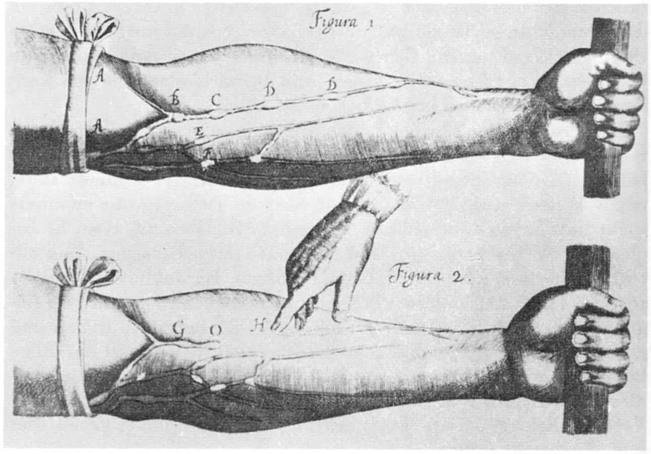
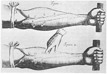 Image of veins from William Harvey's Exercitatio Anatomica de Motu Cordis et Sanguinis in Animalibus
Michael Servetus was the first European to describe the function of pulmonary circulation, although his achievement was not widely recognized at the time, for a few reasons. One was that the description appeared in a theological treatise, Christianismi Restitutio, not in a book on medicine. Most copies of the book were burned shortly after its publication in 1553 because of persecution of Servetus by religious authorities. Finally William Harvey, a pupil of Hieronymus Fabricius (who had earlier described the valves of the veins without recognizing their function), performed a sequence of experiments, and published Exercitatio Anatomica de Motu Cordis et Sanguinis in Animalibus in 1628, which "demonstrated that there had to be a direct connection between the venous and arterial systems throughout the body, and not just the lungs. Most importantly, he argued that the beat of the heart produced a continuous circulation of blood through minute connections at the extremities of the body. This is a conceptual leap that was quite different from Ibn al-Nafis' refinement of the anatomy and bloodflow in the heart and lungs."[10] This work, with its essentially correct exposition, slowly convinced the medical world. However, Harvey was not able to identify the capillary system connecting arteries and veins; these were later discovered by Marcello Malpighi in 1661.
See also
- Microcirculation
- Cardiology
- Lymphatic system
- Blood vessels
- Innate heat
- Cardiac muscle
- Major systems of the human body
- Heart
- Amato Lusitano
- William Harvey
References
- ^ "cardiovascular system" at Dorland's Medical Dictionary
- ^ "circulatory system" at Dorland's Medical Dictionary
- ^ MeSH Cardiovascular+System
- ^ a b Dwivedi, Girish & Dwivedi, Shridhar (2007). History of Medicine: Sushruta – the Clinician – Teacher par Excellence. National Informatics Centre (Government of India).
- ^ Anatomy - History of anatomy
- ^ Mohammadali M. Shojaa, R. Shane Tubbsb, Marios Loukasc, Majid Khalilid, Farid Alakbarlie, Aaron A. Cohen-Gadola; Tubbs, RS; Loukas, M; Khalili, M; Alakbarli, F; Cohen-Gadol, AA (29 May 2009), "Vasovagal syncope in the Canon of Avicenna: The first mention of carotid artery hypersensitivity", International Journal of Cardiology (Elsevier) 134 (3): 297–301, doi:10.1016/j.ijcard.2009.02.035, PMID 19332359
- ^ Rachel Hajar (1999), "The Greco-Islamic Pulse", Heart Views 1 (4): 136-140 [138]
- ^ Chairman's Reflections (2004), "Traditional Medicine Among Gulf Arabs, Part II: Blood-letting", Heart Views 5 (2), p. 74-85 [80].
- ^ West, John B. (October 9, 2008), "Ibn al-Nafis, the pulmonary circulation, and the Islamic Golden Age", Journal of Applied Physiology 105 (6): 1877–80, doi:10.1152/japplphysiol.91171.2008, PMC 2612469, PMID 18845773, http://www.pubmedcentral.nih.gov/articlerender.fcgi?tool=pmcentrez&artid=2612469
- ^ Peter E. Pormann and E. Savage Smith, Medieval Islamic medicine Georgetown University, Washington DC, 2007, p. 48.
External links
- The Circulatory System Article
- The Circulatory System
- NCP Cardiovascular Medicine A Journal Covering Clinical Cardiovascular Medicine
- Reiber C. L. & McGaw I. J. (2009). "A Review of the “Open” and “Closed” Circulatory Systems: New Terminology for Complex Invertebrate Circulatory Systems in Light of Current Findings". International Journal of Zoology 2009: 8 pages. doi:10.1155/2009/301284.
Human systems and organs TA 2–4:
MSBone (Carpus · Collar bone (clavicle) · Thigh bone (femur) · Fibula · Humerus · Mandible · Metacarpus · Metatarsus · Ossicles · Patella · Phalanges · Radius · Skull (cranium) · Tarsus · Tibia · Ulna · Rib · Vertebra · Pelvis · Sternum) · CartilageTA 5–11:
splanchnic/
viscusmostly
Thoracicmostly
AbdominopelvicDigestive system+
adnexaMouth (Salivary gland, Tongue) · upper GI (Oropharynx, Laryngopharynx, Esophagus, Stomach) · lower GI (Small intestine, Appendix, Colon, Rectum, Anus) · accessory (Liver, Biliary tract, Pancreas)TA 12–16 Circulatory systemCardiovascular systemBlood
(Non-TA)General anatomy: systems and organs, regional anatomy, planes and lines, superficial axial anatomy, superficial anatomy of limbsCirculatory system: Arteries and veins (TA A12.0, TH H3.09.02, GA 6.543/GA 7.641) Systemic circulation (Left heart) → Aorta → Arteries → Arterioles → Capillaries → Venules → Veins → Vena cava → (Right heart)Pulmonary circulation Blood vessels Arteries Veins Lymphatic Cardiovascular system, physiology: cardiovascular physiology Heart VolumesStroke volume = End-diastolic volume – End-systolic volume
Cardiac output = Heart rate × Stroke volume
Frank–Starling law of the heart · Cardiac function curve · Venous return curve
Aortic valve area calculation · Ejection fraction · Cardiac indexFractional shortening = (End-diastolic dimension – End-systolic dimension) / End-diastolic dimensionInteraction diagramsChronotropic (Heart rate) · Dromotropic (Conduction velocity) · Inotropic (Contractility) · Bathmotropic (Excitability) · Lusitropic (Relaxation)Cardiac action potential (Atrial action potential, Ventricular action potential) · Effective refractory period · Pacemaker potential · EKG (P wave, PR interval, QRS complex, QT interval, ST segment, T wave, U wave) · Hexaxial reference systemChamber pressureCentral venous pressure/right atrial pressure → Right ventricular pressure → Pulmonary artery pressure → Pulmonary wedge pressure/left atrial pressure → Left ventricular pressure → Aortic pressureOtherVascular system/
HemodynamicsBaroreflex · Kinin-kallikrein system · Renin-angiotensin system · Vasoconstrictors/Vasodilators · Autoregulation (Myogenic mechanism, Tubuloglomerular feedback) · Paraganglia (Aortic body, Carotid body, Glomus cell)Prenatal development/Mammalian development of circulatory system (GA 5, TE E5.11) Heart development Tubular heartSepta/ostiaAtrioventricular cushions/Septum intermedium · Primary interatrial foramen · Septum primum (Foramen secundum) · Septum secundum (Foramen ovale) · Aorticopulmonary septumOtherAtrioventricular canal · Primary interventricular foramenVasculogenesis,
angiogenesis,
and lymphangiogenesisBlood island of umbilical vesicle
Development of arteriesDevelopment of veinsDevelopment of lymph vesselsLymph sacsDevelopment of circulatory system about teeth near childrenanuli: Anulus sanguineus perienameleus · lacunae: Lacuna sanguinea supraenamelea (Ductus sanguineus mesialis · Ductus sanguineus distalis · Ductus sanguineus lingualis · Ductus sanguineus palatinus · Ductus sanguineus buccalis · Ductus sanguineus labialis), Lacuna sanguinea apicalis, Lacuna sanguinea periodontalis, Lacuna sanguinea parodontalis, Lacuna sanguinea gingivalisExtraembryonic
hemangiogenesisFetal circulation Cardiovascular disease: heart disease · Circulatory system pathology (I00–I52, 390–429) Ischaemic Active ischemiaAngina pectoris (Prinzmetal's angina, Stable angina) · Acute coronary (Unstable angina, Myocardial infarction / heart attack)hours (Myocardial stunning, Hibernating myocardium) · days (Myocardial rupture) · weeks (Aneurysm of heart/Ventricular aneurysm, Dressler's syndrome)Layers Infective endocarditis (Subacute bacterial endocarditis) · noninfective endocarditis (Nonbacterial thrombotic endocarditis, Libman-Sacks endocarditis)Valvesmitral (regurgitation, prolapse, stenosis) · aortic (stenosis, insufficiency) · tricuspid (stenosis, insufficiency) · pulmonary (stenosis, insufficiency)Conduction/
arrhythmiaHeart block: Sinoatrial · AV (1°, 2°, 3°) · Intraventricular (Bundle branch/Right/Left, Left anterior fascicular/Left posterior fascicular, Bifascicular/Trifascicular) · Adams–Stokes syndromePremature contractionWolff-Parkinson-White · Lown-Ganong-LevineFlutter/fibrillationPacemakerWandering pacemaker · Ectopic pacemaker/Ectopic beat · Parasystole · Multifocal atrial tachycardia · Pacemaker syndromeOther/ungroupedhexaxial reference system (Right axis deviation, Left axis deviation) · QT (Short QT syndrome) · T (T wave alternans) · ST (Osborn wave, ST elevation, ST depression)Cardiomegaly Other Lymphatic system - Lymphatic trunks and ducts (TA A12.4.01, GA 8.690) Path details ... Blood → Interstitial fluid → Lymph →
Lymph capillary → Afferent lymph vessel (Lymphatic vessel) → Lymph node → Efferent lymph vessel (Lymphatic vessel) →
Lymph trunk (Subclavian lymph trunk • Jugular lymph trunk • Bronchomediastinal lymph trunk • Intestinal lymph trunk → Cisterna chyli • Lumbar lymph trunk → Cisterna chyli) → Lymph duct {Right lymphatic duct and Thoracic duct (left side)} →
Subclavian vein (right and left) → Blood ...Other concepts M: LMO
anat(h, u, t, a, l)/phys/depv
noco/cong/tumr
proc
Categories:


Wikimedia Foundation. 2010.