Truncus arteriosus (embryology)

Truncus arteriosus (embryology)

Infobox Embryology
Name = Truncus arteriosus
Latin =
GraySubject = 135
GrayPage = 514


Caption = Heart of human embryo of about fourteen days. (Truncus arteriosis visible at top.)


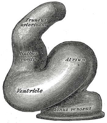
Caption2 = Diagrams to illustrate the transformation of the bulbus cordis. Ao. Truncus arteriosus. Au. Atrium. B. Bulbus cordis. RV. Right ventricle. LV. Left ventricle. P. Pulmonary artery.
System =
CarnegieStage =
Precursor =
GivesRiseTo = aorta, pulmonary artery
MeshName = Truncus+Arteriosus
MeshNumber = A07.541.278.930
DorlandsPre =
DorlandsSuf =
The truncus arteriosus and bulbus cordis are divided by the aorticopulmonary septum.

This makes its appearance in three portions.

(1) Two distal ridge-like thickenings project into the lumen of the tube; these increase in size, and ultimately meet and fuse to form a septum (aorticopulmonary septum), which takes a spiral course toward the proximal end of the truncus arteriosus. It divides the distal part of the truncus into two vessels, the aorta and pulmonary artery, which lie side by side above, but near the heart the pulmonary artery is in front of the aorta.

(2) Four endocardial cushions appear in the proximal part of the truncus arteriosus in the region of the future semilunar valves; the manner in which these are related to the aortic septum is described below.

(3) Two endocardial thickenings—anterior and posterior—develop in the bulbus cordis and unite to form a short septum; this joins above with the aortic septum and below with the ventricular septum. The septum grows down into the ventricle as an oblique partition, which ultimately blends with the ventricular septum in such a way as to bring the bulbus cordis into communication with the pulmonary artery, and through the latter with the sixth pair of aortic arches; while the left ventricle is brought into continuity with the aorta, which communicates with the remaining aortic arches.

Developmental anomalies

Failure of the truncus arteriosus to close results in the condition known a Persistent Truncus Arteriosus. One of cyanotic heart defects or congenital heart defects.


=Additional

External links

*
* [http://sprojects.mmi.mcgill.ca/embryology/cvs/early_chambers.html Overview at mcgill.ca]
* [http://www.med.umich.edu/lrc/coursepages/M1/embryology/embryo/13cardiovascular_system.htm Description and diagram at umich.edu]


Wikimedia Foundation. 2010.

Игры ⚽ Нужен реферат?

Look at other dictionaries:

  • Truncus arteriosus — may refer to:*Persistent truncus arteriosus, a rare congenital heart disease *Truncus arteriosus (embryology), part of an embryo s developing circulatory system …   Wikipedia

  • Embryology — 1 morula, 2 blastula 1 blastula, 2 gastrula with blastopor …   Wikipedia

  • Ductus arteriosus — Artery: Ductus arteriosus Plan of the fetal circulation. ( Ductus arteriosus visible at upper right.) …   Wikipedia

  • human embryology — ▪ biology Introduction       the process encompassing the period from the formation of an embryo, through the development of a fetus, to birth.       The human body, like that of most animals, develops from a single cell produced by the union of… …   Universalium

  • Congenital heart defect — Classification and external resources The normal structure of the heart (left) in comparison to two common locations for a ventricular septal defect (right), the most common form of congenital heart defect.[1] …   Wikipedia

  • Aortic septum — Infobox Embryology Name = PAGENAME Latin = GraySubject = 135 GrayPage = 514 Caption = Heart of human embryo of about thirty five days, opened on right side. (Aortic septum labeled at center right.) Caption2 = Diagrams to show the development of… …   Wikipedia

  • Ductus venosus — Vein: Ductus venosus Fetal circulation. The ductus venosus (red) connects the umbilical vein to the inferior vena cava …   Wikipedia

  • Foramen ovale (heart) — Not to be confused with ostium secundum. Foramen ovale (heart) Sketch showing foramen ovale in a fetal heart. Red arrow shows blood from the inferior caval vein. HF: right atrium, VF: left atrium. HH og VH: right and left ventricle. The heart… …   Wikipedia

  • Cardiale Fehlbildung — Unter einem Herzfehler (auch Herzfehlbildung, Herzvitium) versteht man eine angeborene oder erworbene Strukturbesonderheit des Herzens oder angrenzender Gefäße, die zu Funktionseinschränkungen des Herz Kreislauf Systems oder des Herz Lungen… …   Deutsch Wikipedia

  • Herzfehlbildung — Unter einem Herzfehler (auch Herzfehlbildung, Herzvitium) versteht man eine angeborene oder erworbene Strukturbesonderheit des Herzens oder angrenzender Gefäße, die zu Funktionseinschränkungen des Herz Kreislauf Systems oder des Herz Lungen… …   Deutsch Wikipedia

Share the article and excerpts

Direct link
Do a right-click on the link above
and select “Copy Link”