- Ductus venosus
-
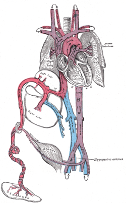
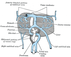
Vein: Ductus venosus Fetal circulation. The ductus venosus (red) connects the umbilical vein to the inferior vena cava. The liver and the veins in connection with it, of a human embryo, twenty-four or twenty-five days old, as seen from the ventral surface. Gray's subject #139 540 Source umbilical vein Drains to inferior vena cava Artery ductus arteriosus In the fetus, the ductus venosus shunts approximately half of the blood flow of the umbilical vein directly to the inferior vena cava. Thus, it allows oxygenated blood from the placenta to bypass the liver. In conjunction with the other fetal shunts, the foramen ovale and ductus arteriosus, it plays a critical role in preferentially shunting oxygenated blood to the fetal brain. It is a part of fetal circulation.
Contents
Postnatal closure
The ductus venosus is open at the time of the birth and is the reason why umbilical vein catheterization works. Ductus venosus naturally closes during the first week of life in most full-term neonates; however, it may take much longer to close in pre-term neonates. Functional closure occurs within minutes of birth. Structural closure in term babies occurs within 3 to 7 days.
After it closes, the remnant is known as ligamentum venosum.
If the ductus venosus fails to occlude after birth, the individual is said to have an intrahepatic portosystemic shunt (PSS). This condition is hereditary in some dog breeds (e.g. Irish Wolfhound). The ductus venosus shows a delayed closure in preterm infants, with no significant correlation to the closure of the ductus arteriosus or the condition of the infant.[1] Possibly, increased levels of dilating prostaglandins leads to a delayed occlusion of the vessel.[1]
See also
References
External links
- 678101050 at GPnotebook - Ductus venosus (embryology)
Prenatal development/Mammalian development of circulatory system (GA 5, TE E5.11) Heart development Tubular heartSepta/ostiaAtrioventricular cushions/Septum intermedium · Primary interatrial foramen · Septum primum (Foramen secundum) · Septum secundum (Foramen ovale) · Aorticopulmonary septumOtherAtrioventricular canal · Primary interventricular foramenVasculogenesis,
angiogenesis,
and lymphangiogenesisBlood island of umbilical vesicle
Development of arteriesDevelopment of veinsDevelopment of lymph vesselsLymph sacsDevelopment of circulatory system about teeth near childrenanuli: Anulus sanguineus perienameleus · lacunae: Lacuna sanguinea supraenamelea (Ductus sanguineus mesialis · Ductus sanguineus distalis · Ductus sanguineus lingualis · Ductus sanguineus palatinus · Ductus sanguineus buccalis · Ductus sanguineus labialis), Lacuna sanguinea apicalis, Lacuna sanguinea periodontalis, Lacuna sanguinea parodontalis, Lacuna sanguinea gingivalisExtraembryonic
hemangiogenesisFetal circulation umbilical cord: Umbilical vein → Ductus venosus → Inferior vena cava → Heart → Pulmonary artery → Ductus arteriosus → Aorta → Umbilical artery
yolk sac: Vitelline veins · Vitelline arteriesVeins of the abdomen and pelvis (TA A12.3.09–10, 12, GA 7.672) To azygos system IVC
(Systemic)To IVC or left renal veininferior phrenic · hepatic (central veins of liver, liver sinusoid) · suprarenal · renal · gonadal (ovarian ♀/testicular ♂, pampiniform plexus ♂) · lumbar · common iliacUnpairedposterior: iliolumbar · superior gluteal · lateral sacral
anterior: inferior gluteal · obturator · uterine ♀ (uterine plexus ♀) · vesical (vesical plexus, prostatic plexus ♂, deep of penis ♂/clitoris ♀, posterior scrotal ♂/labial ♀) · vaginal plexus/vein ♀ · middle rectal · internal pudendal (inferior rectal, bulb of penis ♂/vestibule ♀) · rectal plexusPortal vein
(Portal)DirectCategories:- Embryology of cardiovascular system
- Veins
- Cardiovascular system stubs
Wikimedia Foundation. 2010.


