- Dorsal aorta
-
This article is about the embryonic artery. For the artery in adult fish, see Aorta#In other animals.
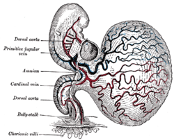
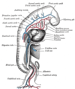
Dorsal aortae Profile view of a human embryo estimated at twenty or twenty-one days old. (Dorsal aorta labeled at center left.) Latin aortae dorsales Gray's subject #135 506 Carnegie stage 9 Code TE E5.11.2.1.3.0.1 Each primitive aorta receives anteriorly a vein—the vitelline vein—from the yolk-sac, and is prolonged backward on the lateral aspect of the notochord under the name of the dorsal aorta.
The dorsal aortæ give branches to the yolk-sac, and are continued backward through the body-stalk as the umbilical arteries to the villi of the chorion.
The two dorsal aortae combine to become the descending aorta in later development.
External links
- Embryology at Temple Heart98/heart97a/sld017
- Embryology at UNSW Notes/git
- cardev-009 — Embryology at UNC
Prenatal development/Mammalian development of circulatory system (GA 5, TE E5.11) Heart development Tubular heartSepta/ostiaAtrioventricular cushions/Septum intermedium · Primary interatrial foramen · Septum primum (Foramen secundum) · Septum secundum (Foramen ovale) · Aorticopulmonary septumOtherAtrioventricular canal · Primary interventricular foramenVasculogenesis,
angiogenesis,
and lymphangiogenesisBlood island of umbilical vesicle
Development of arteriesDevelopment of veinsDevelopment of lymph vesselsLymph sacsDevelopment of circulatory system about teeth near childrenanuli: Anulus sanguineus perienameleus · lacunae: Lacuna sanguinea supraenamelea (Ductus sanguineus mesialis · Ductus sanguineus distalis · Ductus sanguineus lingualis · Ductus sanguineus palatinus · Ductus sanguineus buccalis · Ductus sanguineus labialis), Lacuna sanguinea apicalis, Lacuna sanguinea periodontalis, Lacuna sanguinea parodontalis, Lacuna sanguinea gingivalisExtraembryonic
hemangiogenesisFetal circulation Categories:- Embryology of cardiovascular system
Wikimedia Foundation. 2010.


