- Yolk sac
Infobox Embryology
Name = Yolk sac
Latin =
GraySubject = 11
GrayPage = 54
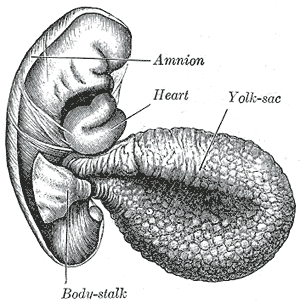
Caption = Humanembryo of 2.6 mm.
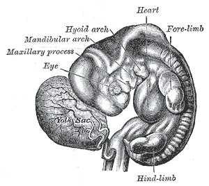
Caption2 = Human embryo from thirty-one to thirty-four days
System =
CarnegieStage = 5b
Days = 9
Precursor =endoderm
GivesRiseTo =
MeshName = Yolk+Sac
MeshNumber = A10.615.284.981
DorlandsPre = s_01
DorlandsSuf = 12716805
The yolk sac is the first element seen in thegestational sac duringpregnancy , usually at 5 weeksgestation .It is a critical landmark, identifying a true gestation sac
It is reliably seen early in pregnancy using
ultrasound .In mice
In the mouse, the yolk sac is the first site of blood formation, generating primitive macrophages and erythrocytes.
In humans
The yolk-sac is situated on the ventral aspect of the
embryo ; it is lined by extra-embyronicendoderm , outside of which is a layer of extra-embryonic mesenchyme, derived from themesoderm .Blood is conveyed to the wall of the sac by the primitive
aorta , and after circulating through a wide-meshed capillary plexus, is returned by thevitelline veins to the tubular heart of the embryo. This constitutes thevitelline circulation , and by means of it nutritive material is absorbed from the yolk-sac and conveyed to the embryo.At the end of the fourth week the yolk-sac presents the appearance of a small pear-shaped vesicle (umbilical vesicle) opening into the digestive tube by a long narrow tube, the
vitelline duct .The vesicle can be seen in the after-birth as a small, somewhat oval-shaped body whose diameter varies from 1 mm. to 5 mm.; it is situated between the
amnion and thechorion and may lie on or at a varying distance from theplacenta .As a rule the duct undergoes complete obliteration during the seventh week, but in about two percent of cases its proximal part persists as a diverticulum from the small intestine,
Meckel's diverticulum , which is situated about 60cm proximal to theileocecal valve , and may be attached by a fibrous cord to the abdominal wall at theumbilicus .Sometimes a narrowing of the lumen of the
ileum is seen opposite the site of attachment of the duct.Histogenesis
The Yolk sac starts forming itself during the second week of the embryonic development, at the same time of the shaping of the amniotic sac. The
hypoblast starts proliferating laterally and descending.In the meantime the Heuser membrane, located on the opposite pole of the developing vesicle, starts its upward proliferation and meets the hypoblast.
Modifications
*Primary yolk sac/primitive yolk sac: it is the vesicle constituted in the second week, its floor is represented by the
Heuser membrane and its ceiling by thehypoblast . It is also known as the exocoelomic cavity. [cite web |url=http://isc.temple.edu/marino/embryology/lecture1-3.htm |title=Text for first three lectures |accessdate=2007-10-13 |format= |work=]*Secondary yolk sac: this first transformation is determined by the modification of its cover, in the connection zone between the ipoblast and the Heuser membrane. We can observe a structure. The two parts detach and the inferior one, which is smaller, forms a cyst destined to be eliminated. The upper one is now covered only by the ipoblast.
*The final yolk sac: during the fourth week of development, during which we can see the shaping of the embryonic areas. A little portion of the sac, in the upper part, constitutes the
intestinal tube . On the other side, the distal part forms a little vesicle that is what remains of the yolk sac.
=Additionalee also
References
Wikimedia Foundation. 2010.