- Umbilical vein
Infobox Vein
Name = Umbilical vein
Latin = vena umbilicalis
GraySubject = 135
GrayPage = 519

Caption =Fetal circulation ; the umbilical vein is the large, red vessel at the far left.
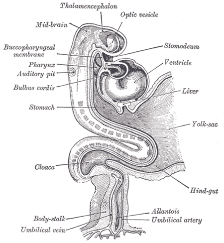
Caption2 = Human embryo about fifteen days old.Brain andheart represented from right side.Digestive tube andyolk sac in median section. (Umbilical vein labeled at bottom right.)
DrainsFrom =
DrainsTo =
Artery =umbilical artery
MeshName =
MeshNumber =
Dorlands = nine/16928421
DorlandsSuf = Umbilical veinsThe umbilical vein is ablood vessel present during fetal development that carriesoxygen atedblood from theplacenta to the growingfetus .Circulation
Attached to the
uterine lining , the placenta is the site ofgas exchange between mother and fetus. The singular umbilical vein carries oxygenated blood from the placenta to the fetus, while two umbilical arteries return deoxygenated blood to the placenta. The three vessels coil around one another within theWharton's jelly of theumbilical cord and enter theabdomen at theumbilicus .Inside the fetus, the vein courses alongside the
falciform ligament and then to theliver 's underside. At thetransverse fissure , the vein divides into two vessels, one larger than the other. The larger of the two is joined by theportal vein , and together they enter the right lobe of the liver. The smaller vessel, now called theductus venosus , diverges away from the liver and joins with theinferior vena cava .Closure
Within a week of birth, the infant's umbilical vein is completely obliterated and is replaced by a fibrous cord called the round ligament of the liver (also called the "ligamentum teres hepatis", from the
Latin meaning the same). It extends from the umbilicus to the transverse fissure, where it joins with theligamentum venosum to separate the left and rightlobe s of the liver.Closure of the umbilical vein usually occurs after the
umbilical arteries have closed. This prolongs the communication between the placenta and fetal heart, allowing for a sort ofautotransfusion of remaining blood from the placenta to the fetus.Recanalization
Under extreme pressure, the round ligament may reopen to allow the passage of blood. Such recanalization is common in patients with
cirrhosis andportal hypertension . Patients with cirrhosis experience rapid growth ofscar tissue in and around the liver, often functionally obstructing nearby vessels. Vessel occlusion increases vascular resistance and therefore leads to hypertension. In portal hypertension, the vessels surrounding theliver are subjected to abnormally high blood pressure—so high, in fact, that the force of the blood pressing against the round ligament is sufficient to recanalize the structure.Catheterization
A newborn baby has a patent umbilical vein for at least a few months. This umbilical vein may be catheterised for ready intravenous access. It may be used as a site for regular transfusion in cases of
erythroblastosis orhemolytic disease .
=AdditionalReferences
* - "Peculiarities in the vascular system of the fetus"
External links
*
See also
*
Human umbilical vein graft
Wikimedia Foundation. 2010.