- Esophagus
-
"Gullet" redirects here. For the African sailboat, see Gulet. For the Dutch soccer coach, see Ruud Gullit."Weasand" redirects here. For other meanings, see Weasand (disambiguation).
Esophagus 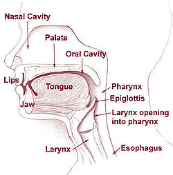
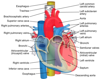
Head and neck. 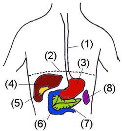
Digestive organs. (Esophagus is #1) Latin œsophagus Gray's subject #245 1144 Artery esophageal arteries Vein esophageal veins Nerve celiac ganglia, vagus[1] Precursor Foregut MeSH oesophagus Dorlands/Elsevier Esophagus The esophagus (or oesophagus) is an organ in vertebrates which consists of a muscular tube through which food passes from the pharynx to the stomach. During swallowing, food passes from the mouth through the pharynx into the esophagus and travels via peristalsis to the stomach. The word esophagus is derived from the Latin œsophagus, which derives from the Greek word oisophagos , lit. "entrance for eating." In humans the esophagus is continuous with the laryngeal part of the pharynx at the level of the C6 vertebra. The esophagus passes through posterior mediastinum in thorax and enters abdomen through a hole in the diaphragm at the level of the tenth thoracic vertebrae (T10). It is usually about 25–30 cm long depending on individual height. It is divided into cervical, thoracic and abdominal parts. Due to the inferior pharyngeal constrictor muscle, the entry to the esophagus opens only when swallowing or vomiting.
Contents
Histology
The layers of the esophagus are as follows:[2]
- mucosa
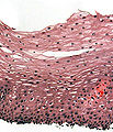
- nonkeratinized stratified squamous epithelium: is rapidly turned over, and serves a protective effect due to the high volume transit of food, saliva and mucus.
- lamina propria: sparse.
- muscularis mucosae: smooth muscle
- submucosa: Contains the mucous secreting glands (esophageal glands), and connective structures termed papillae.
- muscularis externa (or "muscularis propria"): composition varies in different parts of the esophagus, to correspond with the conscious control over swallowing in the upper portions and the autonomic control in the lower portions:
- upper third, or superior part: striated muscle
- middle third, smooth muscle and striated muscle
- inferior third: predominantly smooth muscle
- adventitia
Esophageal constrictions
Normally, the esophagus has three anatomic constrictions at the following levels;[3][4]
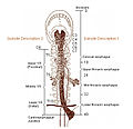
- At the esophageal inlet, where the pharynx joins the esophagus, behind the cricoid cartilage (14-16 cm from the incisor teeth).
- Where its anterior surface is crossed by the aortic arch and the left bronchus (25-27 cm from the incisor teeth).
- Where it pierces the diaphragm (36-38 cm from the incisor teeth).
The distances from the incisor teeth are important as is useful for diagnostic endoscopic procedures.
Gastroesophageal junction
The junction between the esophagus and the stomach (the gastroesophageal junction or GE junction) is not actually considered a valve, although it is sometimes called the cardiac sphincter, cardia or cardias, it actually better resembles a structure.
In much of the gastrointestinal tract, smooth muscles contract in sequence to produce a peristaltic wave which forces a ball of food (called a bolus) while in the esophagus. In humans, peristalsis is found in the contraction of smooth muscles to propel contents through the digestive tract.
In other animals
In most fish, the esophagus is extremely short, primarily due to the length of the pharynx (which is associated with the gills). However, some fish, including lampreys, chimaeras, and lungfish, have no true stomach, so that the esophagus effectively runs from the pharynx directly to the intestine, and is therefore somewhat longer.[5]
In tetrapods, the pharynx is much shorter, and the esophagus correspondingly longer, than in fish. In amphibians, sharks and rays, the esophageal epithelium is ciliated, helping to wash food along, in addition to the action of muscular peristalsis. In the majority of vertebrates, the esophagus is simply a connecting tube, but in birds, it is extended towards the lower end to form a crop for storing food before it enters the true stomach.[5]
A structure with the same name is often found in invertebrates, including molluscs and arthropods, connecting the oral cavity with the stomach.
See also
Additional images
-
H&E stain of biopsy of normal esophagus showing the stratified squamous cell epithelium
References
- ^ Physiology at MCG 6/6ch2/s6ch2_30
- ^ Histology at BU 10801loa
- ^ Snell, Richard (2007). Clinical anatomy by regions. Lippincott Williams & Wilkins. p. 129. ISBN 9780781764049. http://books.google.com/books?id=7SZWRe2OBlgC&pg=PA129.
- ^ Schünke, Michael; Schulte, Erik; Schumacher, Udo; Ross, Lawrence; Lamperti, Edward (2006). Atlas of anatomy: neck and internal organs. Thieme. p. 70. ISBN 9781588904430. http://books.google.com/books?id=n0jqG0Lv2CAC&pg=PA70.
- ^ a b Romer, Alfred Sherwood; Parsons, Thomas S. (1977). The Vertebrate Body. Philadelphia, PA: Holt-Saunders International. pp. 344–345. ISBN 0-03-910284-X.
External links
- Virtual Slidebox at Univ. Iowa Slide 449
- Esophagus Foreign Body MedPix Radiology Teaching File
Human systems and organs TA 2–4:
MSBone (Carpus · Collar bone (clavicle) · Thigh bone (femur) · Fibula · Humerus · Mandible · Metacarpus · Metatarsus · Ossicles · Patella · Phalanges · Radius · Skull (cranium) · Tarsus · Tibia · Ulna · Rib · Vertebra · Pelvis · Sternum) · CartilageTA 5–11:
splanchnic/
viscusmostly
Thoracicmostly
AbdominopelvicDigestive system+
adnexaMouth (Salivary gland, Tongue) · upper GI (Oropharynx, Laryngopharynx, Esophagus, Stomach) · lower GI (Small intestine, Appendix, Colon, Rectum, Anus) · accessory (Liver, Biliary tract, Pancreas)TA 12–16 Blood
(Non-TA)General anatomy: systems and organs, regional anatomy, planes and lines, superficial axial anatomy, superficial anatomy of limbs - mucosa
Wikimedia Foundation. 2010.

