- Serous membrane
Infobox Anatomy
Name = PAGENAME
Latin = tunica serosa
GraySubject =
GrayPage =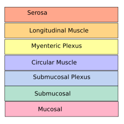
Caption = Layers of theenteric nervous system . (Serosa at top, in red.)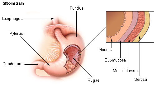
Caption2 = Stomach. (Serosa is labeled at far right, and is colored yellow.)
Precursor =mesoderm
System =
MeshName = Serous+membrane
MeshNumber = A10.615.789
DorlandsPre = t_22
DorlandsSuf = 12832289In
anatomy , a serous membrane (or serosa) is a smooth membrane consisting of a thin layer of cells which excreteserous fluid . Serous membranes line and enclose several body cavities, known as serous cavities, where they secrete a lubricating fluid which reduces friction from muscle movement. Serosa is not to be confused withadventitia , a connective tissue layer which binds together structures rather than reducing friction between them.tructure
Each serous membrane is composed of a secretory epithelial layer and a
connective tissue layer underneath.
* The "epithelial layer", known asmesothelium , consists of a single layer of avascular flatnucleated cells (simple squamous epithelium ) which produce the lubricating serous fluid. This fluid has a consistency similar to thin mucus. These cells are bound tightly to the underlying connective tissue.
* The "connective tissue layer" provides theblood vessels andnerves for the overlying secretory cells, and also serves as the binding layer which allows the whole serous membrane to adhere to organs and other structures.For the heart, the surrounding serous membranes include:
Other parts of the body may also have specific names for these structures. For example, the serosa of the
uterus is called theperimetrium .The pericardial cavity (surrounding the
heart ), pleural cavity (surrounding thelung s) and peritoneal cavity (surrounding most organs of theabdomen ) are the three serous cavities within the human body. It should be noted that while serous membranes have a lubricative role to play in all three cavities, in the pleural cavity it has a greater role to play in the function of breathing.The serous cavities are formed from the intraembryonic coelom and are basically an empty space within the body surrounded by serous membrane. Early in embryonic life visceral organs develop adjacent to a cavity and invaginate into the bag-like coelom. Therefore each organ becomes surrounded by serous membrane - they "do not" lie within the serous cavity. The layer in contact with the organ is known as the visceral layer, while the parietal layer is in contact with the body wall.
Embryological origins
All serous membranes found in the human body formed ultimately from the
mesoderm of thetrilaminar embryo . The trilaminar embryo consists of three relatively flat layers ofectoderm ,endoderm (also known as "entoderm") andmesoderm .As the embryo develops, the mesoderm starts to segment into three main regions: the
paraxial mesoderm , theintermediate mesoderm and thelateral plate mesoderm .The lateral plate mesoderm later splits in half to form two layers bounding a cavity known as the
intraembryonic coelom . Collectively, both layers are known assplanchnopleure . Individually, each are known asvisceropleure andsomatopleure .
* The "visceropleure" is associated with the underlying endoderm which it is in contact with, and later becomes the serous membrane in contact with visceral organs within the body.
* The "somatopleure" is associated with the overlying ectoderm and later becomes the serous membrane in contact with the body wall.The intraembronic coelom can now be seen as a cavity within the body which is covered with serous membrane derived from the splanchnopleure. This cavity is divided and demarcated by the folding and development of the embryo, ultimately forming the serous cavities which house many different organs within the
thorax andabdomen .
=AdditionalReferences
External links
*
* - "Tissues, Layers, and Organs: transverse section of rat gut"
* - "Uterus "
* - "Jejunum "
*
Wikimedia Foundation. 2010.