- Human gastrointestinal tract
-
"Alimentary canal" redirects here. For animal alimentary canal in general, see Gut (anatomy).
Human gastrointestinal tract (Digestive System) 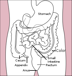
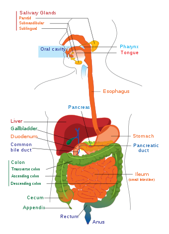
Stomach colon rectum diagram The human gastrointestinal tract refers to the stomach and intestine,[1] and sometimes to all the structures from the mouth to the anus.[2] (The "digestive system" is a broader term that includes other structures, including the accessory organs of digestion).[3]
In an adult male human, the gastrointestinal (GI) tract is 5 metres (20 ft) long in a live subject, or up to 9 metres (30 ft) without the effect of muscle tone, and consists of the upper and lower GI tracts. The tract may also be divided into foregut, midgut, and hindgut, reflecting the embryological origin of each segment of the tract.
The GI tract always releases hormones to help regulate the digestion process. These hormones, including gastrin, secretin, cholecystokinin, and grehlin, are mediated through either intracrine or autocrine mechanisms, indicating that the cells releasing these hormones are conserved structures throughout evolution.[4]
Contents
Upper gastrointestinal tract
 Upper and Lower human gastrointestinal tract
The upper gastrointestinal tract consists of the esophagus, stomach, and duodenum.[5] The exact demarcation between "upper" and "lower" can vary. Upon gross dissection, the duodenum may appear to be a unified organ, but it is often divided into two parts based upon function, arterial supply, or embryology.
Lower gastrointestinal tract
The lower gastrointestinal tract includes most of the small intestine and all of the large intestine.[6] According to some sources, it also includes the anus present in human body.[citation needed]
- Bowel or intestine
- Small intestine, which has three parts:
- Duodenum - Here the digestive juices from pancreas (digestive enzymes) and gallbladder (bile) mix together. The digestive enzymes break down proteins and bile emulsifies fats into micelles. Duodenum contains Brunner's glands which produce bicarbonate and pancreatic juice contains bicarbonate to neutralize hydrochloric acid of stomach
- Jejunum - It is the midsection of the intestine, connecting duodenum to ileum. Contain plicae circulares, and villi to increase surface area.
- Ileum - It has villi, where all soluble molecules are absorbed into the blood (capillaries and lacteals).
- Large intestine, which has three parts:
- Cecum (the vermiform appendix is attached to the cecum).
- Colon (ascending colon, transverse colon, descending colon and sigmoid flexure). The main function of colon is to absorb water, but it also contains bacteria that produce beneficial vitamins like Vitamin K.
- Rectum in human body
- Small intestine, which has three parts:
- Anus
The ligament of Treitz is sometimes used to divide the upper and lower GI tracts.[7]
Embryology
The gut is an endoderm-derived structure. At approximately the sixteenth day of human development, the embryo begins to fold ventrally (with the embryo's ventral surface becoming concave) in two directions: the sides of the embryo fold in on each other and the head and tail fold toward one another. The result is that a piece of the yolk sac, an endoderm-lined structure in contact with the ventral aspect of the embryo, begins to be pinched off to become the primitive gut. The yolk sac remains connected to the gut tube via the vitelline duct. Usually this structure regresses during development; in cases where it does not, it is known as Meckel's diverticulum.
During fetal life, the primitive gut can be divided into three segments: foregut, midgut, and hindgut. Although these terms often are used in reference to segments of the primitive gut, they nevertheless are used regularly to describe components of the definitive gut as well.
Each segment of the gut gives rise to specific gut and gut-related structures in later development. Components derived from the gut proper, including the stomach and colon, develop as swellings or dilatations of the primitive gut. In contrast, gut-related derivatives—that is, those structures that derive from the primitive gut, but are not part of the gut proper—in general develop as outpouchings of the primitive gut. The blood vessels supplying these structures remain constant throughout development.[8]
Part Part in adult Gives rise to Arterial supply Foregut Esophagus to first 2 sections of the duodenum Esophagus, Stomach, Duodenum (1st and 2nd parts), Liver, Gallbladder, Pancreas, Spleen, Superior portion of pancreas celiac trunk Midgut lower duodenum, to the first two-thirds of the transverse colon lower duodenum, jejunum, ileum, cecum, appendix, ascending colon, and first two-thirds of the transverse colon branches of the superior mesenteric artery Hindgut last third of the transverse colon, to the upper part of the anal canal last third of the transverse colon, descending colon, rectum, and upper part of the anal canal branches of the inferior mesenteric artery Transit time
The time taken for food or other ingested objects to transit through the gastrointestinal tract varies depending on many factors, but roughly, it takes 2.5 to 3 hours after a meal for 50% of stomach contents to empty into the intestines and total emptying of the stomach takes 4 to 5 hours. Subsequently, 50% emptying of the small intestine takes 2.5 to 3 hours. Finally, transit through the colon takes 30 to 40 hours.[9]
Pathology
Main article: Digestive diseaseThere are a number of diseases and conditions affecting the gastrointestinal system, including:
- Cancer
- Cholera
- Colorectal cancer
- Diverticulitis
- Enteric duplication cyst
- Gastroenteritis, also known as "stomach flu"; an inflammation of the stomach and intestines
- Giardiasis
- Inflammatory bowel disease (including Crohn's disease and ulcerative colitis)
- Irritable bowel syndrome
- Pancreatitis
- Peptic ulcer disease
- Appendicitis
- Celiac Disease
- Ulcerative colitis
- Yellow Fever
Immune function
The gastrointestinal tract is also a prominent part of the immune system.[10] The surface area of the digestive tract is estimated to be the surface area of a football field. With such a large exposure, the immune system must work hard to prevent pathogens from entering into blood and lymph.[11]
The low pH (ranging from 1 to 4) of the stomach is fatal for many microorganisms that enter it. Similarly, mucus (containing IgA antibodies) neutralizes many of these microorganisms. Other factors in the GI tract help with immune function as well, including enzymes in saliva and bile. Enzymes such as Cyp3A4, along with the antiporter activities, also are instrumental in the intestine's role of detoxification of antigens and xenobiotics, such as drugs, involved in first pass metabolism.
Health-enhancing intestinal bacteria serve to prevent the overgrowth of potentially harmful bacteria in the gut. These two types of bacteria compete for space and "food," as there are limited resources within the intestinal tract. A ratio of 80-85% beneficial to 15-20% potentially harmful bacteria generally is considered normal within the intestines. Microorganisms also are kept at bay by an extensive immune system comprising the gut-associated lymphoid tissue (GALT).
Histology
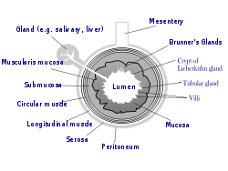 General structure of the gut wall
The gastrointestinal tract has a form of general histology with some differences that reflect the specialization in functional anatomy.[12] The GI tract can be divided into four concentric layers:
- Mucosa
- Submucosa
- Muscularis externa (the external muscular layer)
- Adventitia or serosa
Mucosa
The mucosa is the innermost layer of the gastrointestinal wall that is surrounding the lumen, or open space within the tube. This layer comes in direct contact with food called bolus, and is responsible for absorption, digestion and secretion which are the important processes in digestion.
The mucosa is made up of three layers:
- mucous epithelium - an inner layer
- lamina propria - a layer of connective tissue
- muscularis mucosae - a thin layer of smooth muscle
The mucosae are highly specialized in each organ of the gastrointestinal tract, facing a low pH in the stomach, absorbing a multitude of different substances in the small intestine, and also absorbing specific quantities of water in the large intestine. Reflecting the varying needs of these organs, the structure of the mucosa can consist of invaginations of secretory glands (e.g. gastric pits), or it can be folded in order to increase surface area (in the small intestine, particularly the ileum).
Submucosa
The submucosa consists of a dense irregular layer of connective tissue with large blood vessels, lymphatics, and nerves branching into the mucosa and muscularis externa. It contains Meissner's plexus, an enteric nervous plexus, situated on the inner surface of the muscularis externa.
Muscularis externa
The muscularis externa consists of an inner circular layer and a longitudinal outer muscular layer. The circular muscle layer prevents food from traveling backward and the longitudinal layer shortens the tract. The coordinated contractions of these layers is called peristalsis and propels the bolus, or balled-up food, through the GI tract.
Between the two muscle layers are the myenteric or Auerbach's plexus.
Adventitia
The adventitia consists of several layers of connective tissue.
When the adventitia is facing the mesentery or peritoneal fold, the adventitia is covered by a mesothelium supported by a thin connective tissue layer, together forming a serosa, or serous membrane.
The parts of alimentary canal that are lined by adventitia are oral cavity, esophagus and anal canal.
See also
- Dysbiosis
- Gastrointestinal hormone
- Major systems of the human body
- Mannan Oligosaccharide based nutritional supplements
References
- ^ "gastrointestinal tract" at Dorland's Medical Dictionary
- ^ MeSH Gastrointestinal+tract
- ^ "digestive system" at Dorland's Medical Dictionary
- ^ Nelson RJ. 2005. Introduction to Behavioral Endocrinology. Sinauer Associates: Massachusetts. p 57.
- ^ MeSH Upper+Gastrointestinal+Tract
- ^ MeSH Lower+Gastrointestinal+Tract
- ^ David A. Warrell (2005). Oxford textbook of medicine: Sections 18-33. Oxford University Press. pp. 511–. ISBN 9780198569787. http://books.google.com/books?id=hL1NKQJlY1IC&pg=PA511. Retrieved 1 July 2010.
- ^ Bruce M. Carlson (2004). Human Embryology and Developmental Biology (3rd ed.). Saint Louis: Mosby. ISBN 0-323-03649-X.
- ^ Colorado State University > Gastrointestinal Transit: How Long Does It Take? Last updated on May 27, 2006. Author: R. Bowen.
- ^ Richard Coico, Geoffrey Sunshine, Eli Benjamini (2003). Immunology: a short course. New York: Wiley-Liss. ISBN 0-471-22689-0.
- ^ Animal Physiology textbook
- ^ Abraham L. Kierszenbaum (2002). Histology and cell biology: an introduction to pathology. St. Louis: Mosby. ISBN 0-323-01639-1.
External links
- Pediatric Overview of Human Digestive System
- Anatomy atlas of the Digestive System
- Overview at Colorado State University
- Your Digestive System and How It Works at National Institutes of Health
Human systems and organs TA 2–4:
MSBone (Carpus · Collar bone (clavicle) · Thigh bone (femur) · Fibula · Humerus · Mandible · Metacarpus · Metatarsus · Ossicles · Patella · Phalanges · Radius · Skull (cranium) · Tarsus · Tibia · Ulna · Rib · Vertebra · Pelvis · Sternum) · CartilageTA 5–11:
splanchnic/
viscusmostly
Thoracicmostly
AbdominopelvicDigestive system+
adnexaMouth (Salivary gland, Tongue) · upper GI (Oropharynx, Laryngopharynx, Esophagus, Stomach) · lower GI (Small intestine, Appendix, Colon, Rectum, Anus) · accessory (Liver, Biliary tract, Pancreas)TA 12–16 Blood
(Non-TA)General anatomy: systems and organs, regional anatomy, planes and lines, superficial axial anatomy, superficial anatomy of limbsHead and neck anatomy, digestive system: Mouth anatomy (TA A05.1–2, TH H3.04.01, GA 11.1110–2, 1125–1141) Mouth Lip (Upper, Lower, Vermilion border, Frenulum of lower lip, Labial commissure of mouth, Philtrum)
Cheek (Buccal fat pad)Interdental papilla · Gingival sulcus · Gingival margin · Free gingival margin · Gingival fibers · Junctional epithelium · Mucogingival junction · Sulcular epithelium · Stippling
Periodontium: Cementum · Gingiva · Periodontal ligamentTeethsee tooth anatomydorsum (Taste bud, Median sulcus, Terminal sulcus, Foramen cecum, Lingual tonsils) · underside (Frenulum, Fimbriated fold, Sublingual caruncle) · Anterior · Posterior · Glossoepiglottic folds · Lingual septumOro-pharynx/
faucesCategories:- Digestive system
- Abdomen
- Routes of administration
- Bowel or intestine


Wikimedia Foundation. 2010.