- Major duodenal papilla
-
Major duodenal papilla 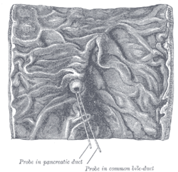
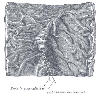
Interior of the descending portion of the duodenum, showing bile papilla. 
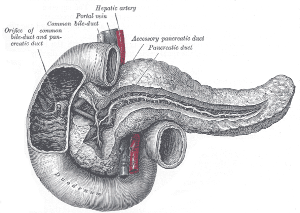
The pancreatic duct. Latin papilla duodeni major  1. Bile ducts: 2. Intrahepatic bile ducts, 3. Left and right hepatic ducts, 4. Common hepatic duct, 5. Cystic duct, 6. Common bile duct, 7. Ampulla of Vater, 8. Major duodenal papilla
9. Gallbladder, 10-11. Right and left lobes of liver. 12. Spleen.
13. Esophagus. 14. Stomach. Small intestine: 15. Duodenum, 16. Jejunum
17. Pancreas: 18: Accessory pancreatic duct, 19: Pancreatic duct.
20-21: Right and left kidneys (silhouette).
The anterior border of the liver is lifted upwards (brown arrow). Gallbladder with Longitudinal section, pancreas and duodenum with frontal one. Intrahepatic ducts and stomach in transparency.The common bile duct and the pancreatic duct together perforate the medial side of the second portion of the duodenum obliquely, some 7 to 10 cm below the pylorus, forming a structure called the major duodenal papilla.
The accessory pancreatic duct sometimes pierces it about 2 cm above and slightly in front of these.
See also
External links
- pancreas at The Anatomy Lesson by Wesley Norman (Georgetown University)
- digest-019 — Embryology at UNC
- Swiss embryology (from UL, UB, and UF) sdigestive/pankreas01
This article was originally based on an entry from a public domain edition of Gray's Anatomy. As such, some of the information contained within it may be outdated.
Categories:- Digestive system
- Digestive system stubs

Wikimedia Foundation. 2010.