- Human skin
-
Skin 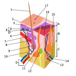
epidermis (A), dermis (B), and subcutis (C), showing a hair follicle (1), sweat gland (11) & sebaceous gland (7) Latin cutis Code TH H3.12.00.1.00001 The human skin is the outer covering of the body. In humans, it is the largest organ of the integumentary system. The skin has multiple layers of ectodermal tissue and guards the underlying muscles, bones, ligaments and internal organs.[1] Human skin is similar to that of most other mammals, except that it is not protected by a pelt. Though nearly all human skin is covered with hair follicles, it appears hairless. There are two general types of skin, hairy and glabrous skin.[2] The adjective cutaneous literally means "of the skin" (from Latin cutis, skin).
Because it interfaces with the environment, skin plays a key role in protecting (the body) against pathogens[3] and excessive water loss.[4] Its other functions are insulation, temperature regulation, sensation, synthesis of vitamin D, and the protection of vitamin B folates. Severely damaged skin will try to heal by forming scar tissue. This is often discolored and depigmented.
In humans, skin pigmentation varies among populations, and skin type can range from dry to oily. Such skin variety provides a rich and diverse habit for bacteria which number roughly at 1000 species from 19 phyla.[5][6]
Contents
Skin components
Skin has mesodermal cells, pigmentation, or melanin provided by melanocytes, which absorb some of the potentially dangerous ultraviolet radiation (UV) in sunlight. It also contains DNA-repair enzymes that help reverse UV damage, and people who lack the genes for these enzymes suffer high rates of skin cancer. One form predominantly produced by UV light, malignant melanoma, is particularly invasive, causing it to spread quickly, and can often be deadly. Human skin pigmentation varies among populations in a striking manner. This has led to the classification of people(s) on the basis of skin color.[7]
The skin is the largest organ in the human body. For the average adult human, the skin has a surface area of between 1.5-2.0 square metres (16.1-21.5 sq ft.), most of it is between 2–3 mm (0.10 inch) thick. The average square inch (6.5 cm²) of skin holds 650 sweat glands, 20 blood vessels, 60,000 melanocytes, and more than 1,000 nerve endings.
Functions
Skin performs the following functions:
- Protection: an anatomical barrier from pathogens and damage between the internal and external environment in bodily defense; Langerhans cells in the skin are part of the adaptive immune system.[3][4]
- Sensation: contains a variety of nerve endings that react to heat and cold, touch, pressure, vibration, and tissue injury; see somatosensory system and haptics.
- Heat regulation: the skin contains a blood supply far greater than its requirements which allows precise control of energy loss by radiation, convection and conduction. Dilated blood vessels increase perfusion and heatloss, while constricted vessels greatly reduce cutaneous blood flow and conserve heat.
- Control of evaporation: the skin provides a relatively dry and semi-impermeable barrier to fluid loss.[4] Loss of this function contributes to the massive fluid loss in burns.
- Aesthetics and communication: others see our skin and can assess our mood, physical state and attractiveness.
- Storage and synthesis: acts as a storage center for lipids and water, as well as a means of synthesis of vitamin D by action of UV on certain parts of the skin.
- Excretion: sweat contains urea, however its concentration is 1/130th that of urine, hence excretion by sweating is at most a secondary function to temperature regulation.
- Absorption: the cells comprising the outermost 0.25–0.40 mm of the skin are "almost exclusively supplied by external oxygen", although the "contribution to total respiration is negligible".[8] In addition, medicine can be administered through the skin, by ointments or by means of adhesive patch, such as the nicotine patch or iontophoresis. The skin is an important site of transport in many other organisms.
- Water resistance: The skin acts as a water resistant barrier so essential nutrients aren't washed out of the body.
Pigments
There are at least five different pigments that determine the color of the skin[9][10]. These pigments are present at different levels and places.
- Melanin: It is brown in color and present in the germinative zone of the epidermis.
- Melanoid: It resembles melanin but is present diffusely throughout the epidermis.
- Carotene: This pigment is yellow to orange in color. It is present in the stratum corneum and fat cells of dermis and superficial fascia.
- Hemoglobin (also spelled haemoglobin): It is found in blood and is not a pigment of the skin but develops a purple color.
- Oxyhemoglobin: It is also found in blood and is not a pigment of the skin. It develops a red color.
Hygiene and skin care
The skin supports its own ecosystems of microorganisms, including yeasts and bacteria, which cannot be removed by any amount of cleaning. Estimates place the number of individual bacteria on the surface of one square inch (6.5 square cm) of human skin at 50 million, though this figure varies greatly over the average 20 square feet (1.9 m2) of human skin. Oily surfaces, such as the face, may contain over 500 million bacteria per square inch (6.5 cm²). Despite these vast quantities, all of the bacteria found on the skin's surface would fit into a volume the size of a pea.[11] In general, the microorganisms keep one another in check and are part of a healthy skin. When the balance is disturbed, there may be an overgrowth and infection, such as when antibiotics kill microbes, resulting in an overgrowth of yeast. The skin is continuous with the inner epithelial lining of the body at the orifices, each of which supports its own complement of microbes.
Proper skin hygiene is important because unclean skin favors the development of pathogenic organisms. The dead cells that continually slough off the epidermis mix with the secretions of the sweat and sebaceous glands and the dust found on the skin form a filthy layer on its surface. If not washed away, the slurry of sweat and sebaceous secretions mixed with dirt and dead skin is decomposed by bacterial flora, producing a foul smell. Functions of the skin are disturbed when it is excessively dirty; it becomes more easily damaged, the release of antibacterial compounds decreases, and dirty skin is more prone to develop infections.[citation needed]
Cosmetics should be used carefully on the skin because these may cause allergic reactions. Each season requires suitable clothing in order to facilitate the evaporation of the sweat. Sunlight, water and air play an important role in keeping the skin healthy.
Oily skin
Oily skin is caused by over-active sebaceous glands, that produce a substance called sebum, a naturally healthy skin lubricant.[1] When the skin produces excessive sebum, it becomes heavy and thick in texture. Oily skin is typified by shininess, blemishes and pimples.[1] The oily-skin type is not necessarily bad, since such skin is less prone to wrinkling, or other signs of aging,[1] because the oil helps to keep needed moisture locked into the epidermis (outermost layer of skin).
The negative aspect of the oily-skin type is that oily complexions are especially susceptible to clogged pores, blackheads, and buildup of dead skin cells on the surface of the skin.[1] Oily skin can be sallow and rough in texture and tends to have large, clearly visible pores everywhere, except around the eyes and neck.[1]
The goal of treating oily skin is to remove excess surface sebum without complete removal of skin lipids.[1] Severe degreasing treatment can foster an actual worsening of sebum secretion, which defeats the aim of the cleansing.[1] A method of cleansing oily skin is to cleanse with a natural face cleanser formulated especially for oily skin. The cleansers pH should be 4.5 - 5.5, since the skin's pH value is approximately 5.4. Gel cleansers work best on oily skin.[1] (see: surfactant) Oily skin products should contain very little natural oils. They should not contain waxes or other synthetic lipid agents that could aggravate the oily condition of the skin. A toning lotion should also be natural and have a pH of 4.5-5.5[citation needed] and formulated especially to help balance and hydrate oily skin. Some cleansing products have lower concentrations of hydroxy acids, which remove dead cells from the upper levels of the stratum corneum.[1] Those products should be used on a regular basis to work adequately.
Aging
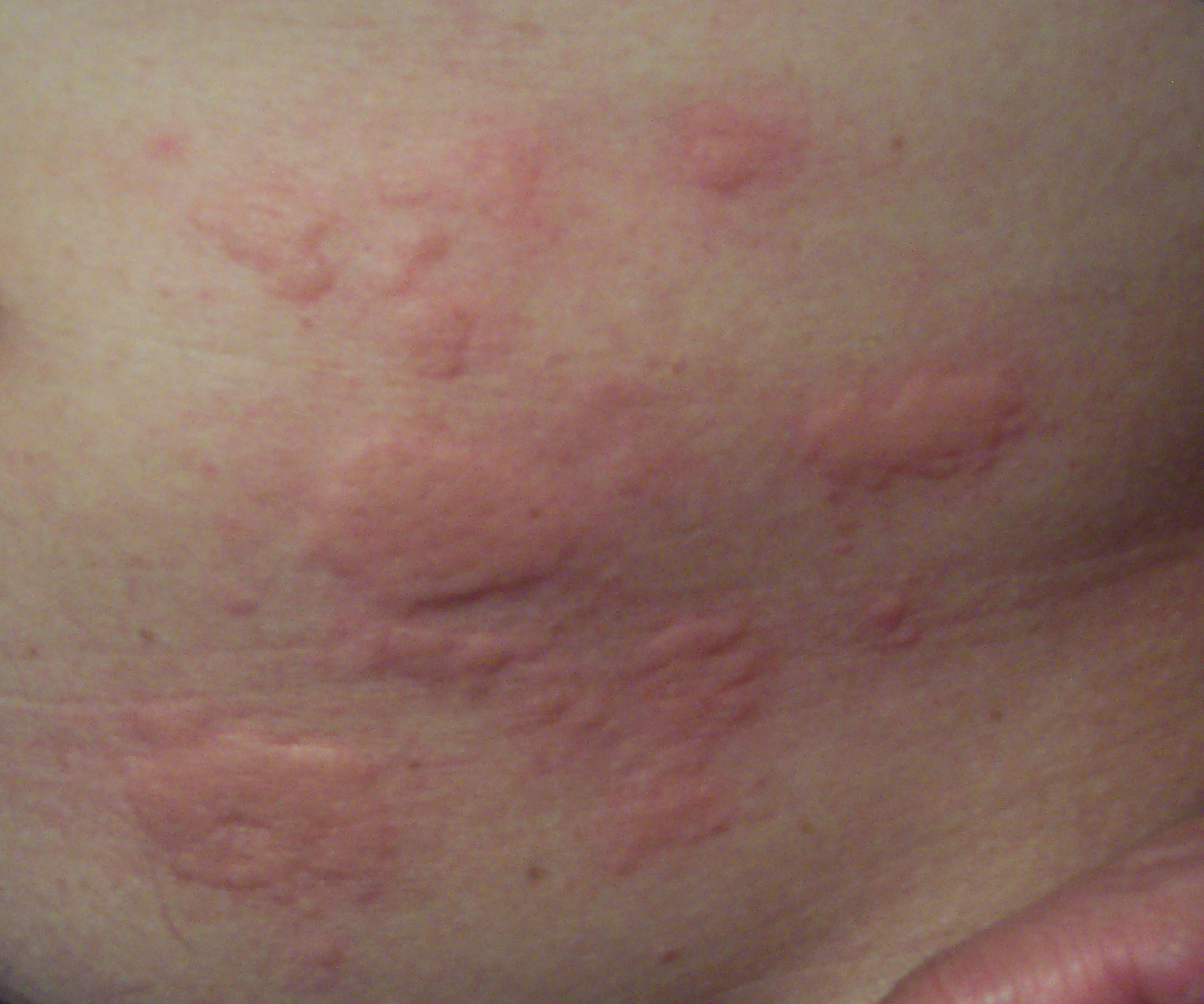
For more details on this topic, see Intrinsic and extrinsic aging.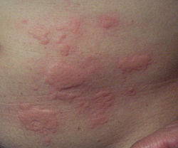 A typical rash
As skin ages, it becomes thinner and more easily damaged. Intensifying this effect is the decreasing ability of skin to heal itself as a person ages.
Among other things, skin aging is noted by a decrease in volume and elasticity. There are many internal and external causes to skin aging. For example: Aging skin receives less blood flow and lower glandular activity.
A validated comprehensive grading scale has categorized the clinical findings of skin aging as laxity (sagging), rhytids (wrinkles), and the various facets of photoaging, including erythema/telangiectasia (redness), dyspigmentation (brown discoloration), solar elastosis (yellowing), keratoses (abnormal growths) and poor texture.[12]
Cortisol causes degradation of collagen,[13] accelerating skin aging.[14]
Photoaging
Photoaging has two main concerns: an increased risk for skin cancer and the appearance of damaged skin. In younger skin, sun damage will heal faster since the cells in the epidermis have a faster turnover rate, while in the older population the skin becomes thinner and the epidermis turnover rate for cell repair is lower which may result in the dermis layer being damaged.[15]
Disease
Diseases of the skin include skin infections and skin neoplasms (including skin cancer).
Dermatology is the branch of medicine that deals with conditions of the skin.[2]
Variability in skin tone
Individuals with ancestors from different parts of the world can have highly visible differences in skin pigmentation. Individuals with sub-Saharan African ancestry (black people) tend towards darker skin, while those of Northern European descent (Nordic people) have paler skin. Between these extremes are individuals of Asian, South-East Asian, Native American, Middle Eastern, Polynesian and Melanesian descent.
The skin of black people has more variation in color from one part of the body to another than does the skin of other racial groups, particularly the palms of the hands and soles of the feet. Part of this is the result of the variations in the thickness of the skin on different parts of the body. The thicker the skin, the more layers of cells with melanin in them, and the darker the color.[16]
Description I Always burns, never tans Pale, Fair, Freckles II Usually burns, sometimes tans Fair III May burn, usually tans Light Brown IV Rarely burns, always tans Olive brown V Moderate constitutional pigmentation Brown VI Marked constitutional pigmentation Black Skin flora
The human skin is a rich environment for microbes.[5][6] Around 1000 species of bacteria from 19 bacterial phyla have been found. Most come from only four phyla: Actinobacteria (51.8%), Firmicutes (24.4%), Proteobacteria (16.5%), and Bacteroidetes (6.3%). Propionibacteria and Staphylococci species were the main species in sebaceous areas. There are three main ecological areas: moist, dry and sebaceous. In moist places on the body Corynebacteria together with Staphylococci dominate. In dry areas, there is a mixture of species but dominated by b-Proteobacteria and Flavobacteriales. Ecologically, sebaceous areas had greater species richness than moist and dry ones. The areas with least similarity between people in species were the spaces between fingers, the spaces between toes, axillae, and umbilical cord stump. Most similarly were beside the nostril, nares (inside the nostril), and on the back.
Reflecting upon the diversity of the human skin researchers on the human skin microbiome have observed: "hairy, moist underarms lie a short distance from smooth dry forearms, but these two niches are likely as ecologically dissimilar as rainforests are to deserts."[5]
The NIH has been launched the Human Microbiome Project to characterize the human microbiota which includes that on the skin and the role of this microbiome in health and disease.[17]
Skin layers
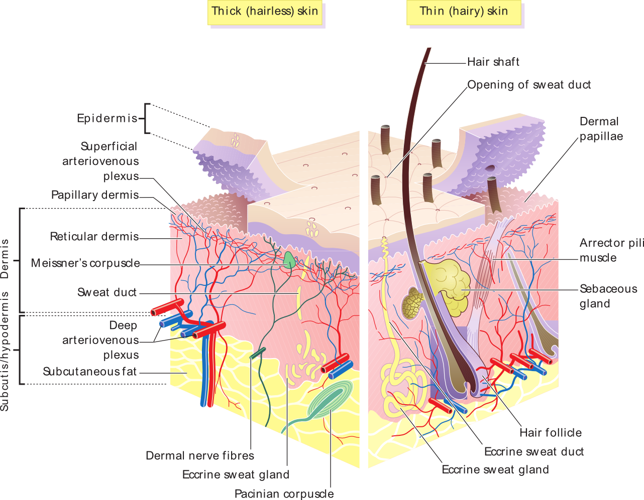
Skin is composed of three primary layers:
- the epidermis, which provides waterproofing and serves as a barrier to infection;
- the dermis, which serves as a location for the appendages of skin; and
- the hypodermis (subcutaneous adipose layer).
Epidermis
Epidermis, "epi" coming from the Greek meaning "over" or "upon", is the outermost layer of the skin. It forms the waterproof, protective wrap over the body's surface and is made up of stratified squamous epithelium with an underlying basal lamina.
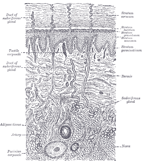
The epidermis contains no blood vessels, and cells in the deepest layers are nourished by diffusion from blood capillaries extending to the upper layers of the dermis. The main type of cells which make up the epidermis are Merkel cells, keratinocytes, with melanocytes and Langerhans cells also present. The epidermis can be further subdivided into the following strata (beginning with the outermost layer): corneum, lucidum (only in palms of hands and bottoms of feet), granulosum, spinosum, basale. Cells are formed through mitosis at the basale layer. The daughter cells (see cell division) move up the strata changing shape and composition as they die due to isolation from their blood source. The cytoplasm is released and the protein keratin is inserted. They eventually reach the corneum and slough off (desquamation). This process is called keratinization and takes place within about 27 days. This keratinized layer of skin is responsible for keeping water in the body and keeping other harmful chemicals and pathogens out, making skin a natural barrier to infection.
 2D projection of a 3D OCT-tomogram of the skin at the fingertip, depicting the stratum corneum (~500 µm thick) with the stratum disjunctum on top and the stratum lucidum in the middle. At the bottom are the superficial parts of the dermis. The sweatducts are clearly visible. (See also: Rotating 3D Version)
Components
The epidermis contains no blood vessels, and is nourished by diffusion from the dermis. The main type of cells which make up the epidermis are keratinocytes, melanocytes, Langerhans cells and Merkels cells. The epidermis helps the skin to regulate body temperature.[citation needed]
Layers
Epidermis is divided into several layers where cells are formed through mitosis at the innermost layers. They move up the strata changing shape and composition as they differentiate and become filled with keratin. They eventually reach the top layer called stratum corneum and are sloughed off, or desquamated. This process is called keratinization and takes place within weeks. The outermost layer of the epidermis consists of 25 to 30 layers of dead cells.
Sublayers
Epidermis is divided into the following 5 sublayers or strata:
- Stratum corneum
- Stratum lucidum
- Stratum granulosum
- Stratum spinosum
- Stratum germinativum (also called "stratum basale")
Mnemonics that are good for remembering the layers of the skin (using "stratum basale" instead of "stratum germinativum"):
- "Can Long Get Some Burgers" (from superficial to deep)
- "Cher Likes Getting Skin Botoxed" (from superficial to deep)
- "Before Signing, Get Legal Counsel" (from deep to superficial)
- "Bare Skin Grows Like Corn" (from deep to superficial)
Blood capillaries are found beneath the epidermis, and are linked to an arteriole and a venule. Arterial shunt vessels may bypass the network in ears, the nose and fingertips.
Skin fields
Dermis 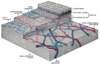
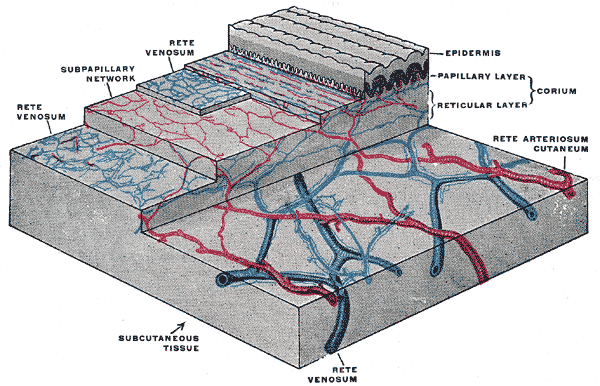
The distribution of the bloodvessels in the skin of the sole of the foot. (Corium - TA alternate term for dermis - is labeled at upper right.) 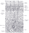
A diagrammatic sectional view of the skin (click on image to magnify). (Dermis labeled at center right.) Gray's subject #234 1065 MeSH Dermis Dorlands/Elsevier Skin Dermis
The dermis is the layer of skin beneath the epidermis that consists of connective tissue and cushions the body from stress and strain. The dermis is tightly connected to the epidermis by a basement membrane. It also harbors many Mechanoreceptor/nerve endings that provide the sense of touch and heat. It contains the hair follicles, sweat glands, sebaceous glands, apocrine glands, lymphatic vessels and blood vessels. The blood vessels in the dermis provide nourishment and waste removal from its own cells as well as from the Stratum basale of the epidermis.
The dermis is structurally divided into two areas: a superficial area adjacent to the epidermis, called the papillary region, and a deep thicker area known as the reticular region.
Papillary region
The papillary region is composed of loose areolar connective tissue. It is named for its fingerlike projections called papillae, that extend toward the epidermis. The papillae provide the dermis with a "bumpy" surface that interdigitates with the epidermis, strengthening the connection between the two layers of skin.
In the palms, fingers, soles, and toes, the influence of the papillae projecting into the epidermis forms contours in the skin's surface. These are called friction ridges, because they help the hand or foot to grasp by increasing friction. Friction ridges occur in patterns (see: fingerprint) that are genetically and epigenetically determined and are therefore unique to the individual, making it possible to use fingerprints or footprints as a means of identification.
Reticular region
The reticular region lies deep in the papillary region and is usually much thicker. It is composed of dense irregular connective tissue, and receives its name from the dense concentration of collagenous, elastic, and reticular fibers that weave throughout it. These protein fibers give the dermis its properties of strength, extensibility, and elasticity.
Also located within the reticular region are the roots of the hair, sebaceous glands, sweat glands, receptors, nails, and blood vessels.
Tattoo ink is held in the dermis. Stretch marks from pregnancy are also located in the dermis.
Hypodermis
The hypodermis is not part of the skin, and lies below the dermis. Its purpose is to attach the skin to underlying bone and muscle as well as supplying it with blood vessels and nerves. It consists of loose connective tissue and elastin. The main cell types are fibroblasts, macrophages and adipocytes (the hypodermis contains 50% of body fat). Fat serves as padding and insulation for the body.
Microorganisms like Staphylococcus epidermidis colonize the skin surface. The density of skin flora depends on region of the skin. The disinfected skin surface gets recolonized from bacteria residing in the deeper areas of the hair follicle, gut and urogenital openings.
Permeability
Human skin permeability is the ability of foreign substances to penetrate and diffuse through the skin. Skin naturally has a low permeability, thus protects the body from particles and foreign toxins by not allowing them to penetrate through the surface. However, technologies in nanomedicine and biology have led to developments in techniques to increase permeability of skin for various applications. The stratum corneum is the outermost layer of skin and is an effective barrier to most inorganic nanosized particles.[18][19] Nanomedicine researchers are interested in nanoparticles that can penetrate the stratum corneum and settle in the epidermis where cells primarily reproduce. If the nanoparticles are targeted to surround cancer cells, they can be used to map where the cancer is located and deliver therapeutic agents directly to the site.
Nanoparticles
Nanoparticles 40 nm in diameter and smaller have been successful in penetrating the skin.[20][21][22] Research confirms that nanoparticles larger than 40 nm do not penetrate the skin past the stratum corneum.[20] Most particles that do penetrate will diffuse through skin cells, but some will travel down hair follicles and reach the dermis layer.
The permeability of skin relative to different shapes of nanoparticles has also been studied. Research has shown that spherical particles have a better ability to penetrate the skin better than oblong (ellipsoidal) particles because spheres are symmetric in all three spacial dimensions.[22] One study compared the two shapes and recorded data that showed spherical particles located deep in the epidermis and dermis whereas ellipsoidal particles were mainly found in the stratum corneum and epidermal layers.[22] Nanorods are used in experiments because of their unique fluorescent properties but have shown mediocre penetration.
Nanoparticles of different materials have shown skin’s permeability limitations. In many experiments, gold nanoparticles 40 nm in diameter or smaller are used and have shown to penetrate to the epidermis. Titanium oxide (TiO2), zinc oxide (ZnO), and silver nanoparticles are ineffective in penetrating the skin past the stratum corneum.[19][23] Cadmium selenide (CdSe) quantum dots have proven to penetrate very effectively when they have certain properties. Because CdSe is toxic to living organisms, the particle must be covered in a surface group. An experiment comparing the permeability of quantum dots coated in polyethylene glycol (PEG), PEG-amine, and carboxylic acid concluded the PEG and PEG-amine surface groups allowed for the greatest penetration of particles. The carboxylic acid coated particles did not penetrate past the stratum corneum.[22]
Increasing permeability
Scientists believed that the skin was an effective barrier to all inorganic particles and alterations to the skin using mechanical stressors causing damage to the skin would be the only way to increase permeability.[24] However, simpler and more effective methods have been developed. For example, ultraviolet radiation (UVR) has been used to slightly damage the surface of skin, causing a time-dependent defect allowing easier penetration of nanoparticles.[25] The UVR’s high energy causes a restructuring of cells, weakening the boundary between the stratum corneum and the epidermal layer.[24][25] The damage of the skin is typically measured by the transepidermal water loss (TEWL), though it may take 3–5 days for the TEWL to reach its peak value. When the TEWL reaches its highest value, the maximum density of nanoparticles is able to permeate the skin. Studies confirm that UVR damaged skin significantly increases the permeability.[24][25] The effects of increased permeability after UVR exposure can lead to an increase in the number of particles that permeate the skin. However, the specific permeability of skin after UVR exposure relative to particles of different sizes and materials has not been determined.[25]
Other skin damaging methods used to increase nanoparticle penetration include tape stripping, skin abrasion, and chemical enhancement. Tape stripping is the process in which tape is applied to skin then lifted to remove the top layer of skin. Skin abrasion is done by shaving the top 5-10 micrometers off the surface of the skin. Chemical enhancement is the process in which chemicals such as polyvinylpyrrolidone (PVP), dimethyl sulfoxide (DMSO), and oleic acid are applied to the surface of the skin to increase permeability.[26][27]
Electroporation is the application of short pulses of electric fields on skin and has proven to increase skin permeability. The pulses are high voltage and on the order of milliseconds when applied. Charged molecules penetrate the skin more frequently than neutral molecules after the skin has been exposed to electric field pulses. Results have shown molecules on the order of 100 micrometers to easily permeate electroporated skin.[27]
Applications
A large area of interest in nanomedicine is the transdermal patch because of the possibility of a painless application of therapeutic agents with very few side effects. Transdermal patches have been limited to administer a small number of drugs, such as nicotine, because of the limitations in permeability of the skin. Development of techniques that increase skin permeability has led to more drugs that can be applied via transdermal patches and more options for patients.[27]
Increasing the permeability of skin allows nanoparticles to penetrate and target cancer cells. Nanoparticles along with multi-modal imaging techniques have been used as a way to diagnose cancer non-invasively. Skin with high permeability allowed quantum dots with an antibody attached to the surface for active targeting to successfully penetrate and identify cancerous tumors in mice. Tumor targeting is beneficial because the particles can be excited using fluorescence microscopy and emit light energy and heat that will destroy cancer cells.[28]
Sunblock and sunscreen
Although some believe that sunblock and sunscreen are both the same, they are not, although they have similar properties and are both important in caring of the skin.
Sunblock Sunblock is opaque and is stronger than sunscreen since it is able to block majority of the UVA/UVB rays and radiation from the sun, thus not having to be reapplied several times a day. Titanium dioxide and zinc oxide are two of the important ingredients in sunblock.
Sunscreen Sunscreen is more transparent once applied to the skin and also has the ability to protect against UVA/UVB rays as well, although the sunscreen's ingredients have the ability to break down at a faster rate once exposed to sunlight, and some of the radiation is able to penetrate to the skin. In order for sunscreen to be more effective it is necessary to consistently reapply and use a higher spf.
Nutrition for healthy skin
Vitamin A, also known as retinoids, benefits the skin by normalizing keratinization, downregulating sebum production which contributes to acne, and reversing and treating photodamage, striae, and cellulite.
Vitamin D and analogs are used to downregulate the cutaneous immune system and epithelial proliferation while promoting differentiation.
Vitamin C is an antioxidant that regulates collagen synthesis, forms barrier lipids, regenerates vitamin E, and provides photoprotection.
Vitamin E is a membrane antioxidant that protects against oxidative damage and also provides protection against harmful UV rays. [29]
Skin overview
 Skin layers, of both hairy and hairless skin
See also
- Acid mantle
- Anthropodermic bibliopegy
- Artificial skin
- Callus, thick area of skin
- List of cutaneous conditions
- Cutaneous structure development
- Fingerprint, skin on fingertips
- Hyperpigmentation, about excess skin color
- Intertriginous
- Meissner's corpuscle
- Pacinian corpuscle
- Polyphenol antioxidant
- Skin lesion
- Skin repair
- Superficial fascia
- Baumann skin types
References
- ^ a b c d e f g h i j "Skin care" (analysis), Health-Cares.net, 2007, webpage: HCcare.
- ^ a b Marks, James G; Miller, Jeffery (2006). Lookingbill and Marks' Principles of Dermatology. (4th ed.). Elsevier Inc. ISBN 1-4160-3185-5.
- ^ a b Proksch, E; Brandner, JM; Jensen, JM (2008). "The skin: an indispensable barrier.". Experimental dermatology 17 (12): 1063–72. PMID 19043850.
- ^ a b c Madison, KC. (2003). "Barrier function of the skin: "la raison d'être" of the epidermis". J Invest Dermatol 121 (2): 231–41. doi:10.1046/j.1523-1747.2003.12359.x. PMID 12880413. http://www.nature.com/jid/journal/v121/n2/pdf/5601872a.pdf.
- ^ a b c Grice, E. A.; Kong, H. H.; Conlan, S.; Deming, C. B.; Davis, J.; Young, A. C.; Bouffard, G. G.; Blakesley, R. W. et al. (2009). "Topographical and Temporal Diversity of the Human Skin Microbiome". Science 324 (5931): 1190–2. doi:10.1126/science.1171700. PMC 2805064. PMID 19478181. http://www.pubmedcentral.nih.gov/articlerender.fcgi?tool=pmcentrez&artid=2805064.
- ^ a b Pappas S. (2009). Your Body Is a Wonderland ... of Bacteria. ScienceNOW Daily News
- ^ Maton, Anthea; Jean Hopkins, Charles William McLaughlin, Susan Johnson, Maryanna Quon Warner, David LaHart, Jill D. Wright (1993). Human Biology and Health. Englewood Cliffs, New Jersey, USA: Prentice Hall. ISBN 0-13-981176-1.
- ^ Stücker, M., A. Struk, P. Altmeyer, M. Herde, H. Baumgärtl & D.W. Lübbers (2002). The cutaneous uptake of atmospheric oxygen contributes significantly to the oxygen supply of human dermis and epidermis.PDF Journal of Physiology 538(3): 985–994. doi:10.1113/jphysiol.2001.013067
- ^ Handbook of General Anatomy by B. D. Chaurasia. ISBN 978-81-239-1654-5
- ^ Pigmentation of Skin
- ^ Theodor Rosebury. Life on Man: Secker & Warburg, 1969 ISBN 0-670-42793-4
- ^ Alexiades-Armenakas, M. R., et al. The spectrum of laser skin resurfacing: nonablative, fractional, and ablative laser resurfacing.J Am Acad Dermatol. 2008 May;58(5):719-37; quiz 738-40
- ^ http://www3.interscience.wiley.com/journal/107640112/abstract
- ^ http://www.ingentaconnect.com/content/bsc/ics/2004/00000026/00000002/art00010
- ^ Gilchrest, BA (1990). "Skin aging and photoaging.". Dermatology nursing / Dermatology Nurses' Association 2 (2): 79–82. PMID 2141531.
- ^ Smith, Wilma; Burns, Catherine (1999). "Managing the hair and skin of African American pediatric patients". Journal of Pediatric Health Care 13 (2): 72–8. doi:10.1016/S0891-5245(99)90057-3. PMID 10382468.
- ^ NIH Human Microbiome Project.
- ^ Baroli, B. Penetration of Nanoparticles and Nanomaterials in the Skin: Fiction or Reality? Journal of Pharmaceutical Sciences 2009 December;99:21-50
- ^ a b Felipe, P., Silva, J.N., Silva, R., Cirne de Castro, J.L., Gomes, M., Alves, L.C., et al. Stratum Corneum Is an Effective Carrier to TiO2 and ZnO Nanoparticle Percutaneous Absorption. Skin Pharmacology and Physiology 2009;22:266-275
- ^ a b Vogt, A., Combadiere, B., Hadam, S., Stieler, K., Lademann, J., Schaefer, H., et al. 40nm, but not 750 or 1,500 nm, Nanoparticles Enter Epidermal CD1a+ Cells after Transcutaneous Application on Human Skin. Journal of Investigative Dermatology
- ^ a b c d Ryman-Rasmussen, J.P., Riviere, J.E. and Monteiro-Riviere, N.A. Penetration of Intact Skin by Quantum Dots with Diverse Physicochemical Properties. Toxicological Sciences 2006;91(1):159-165
- ^ Larese, F., D’Agostin, F., Crosera, M., Adami, G., Renzi, N., Bovenzi, M., et. al. Human skin penetration of silver nanoparticles through intact and damaged skin. Toxicology 2009 September;255:33-37
- ^ a b c Mortensen, L., Oberdorster, G., Pentland, A. and DeLoiuse, L. In Vivo Skin Penetration of Quantum Dot Nanoparticles in the Murine Model: The Effects of UVR. Nano Letters 2008;8(9):2779-2787
- ^ a b c d Mortensen, L., Zheng, H., Faulknor, R., De Benedetto, A., Beck, L., DeLouise, L.A. Increased in vivo skin penetration of quantum dots with UVR and in vitro quantum dot cytotoxicity. Colloidal Quantum Dots for Biomedical Applications IV 2009;7189:1605
- ^ Sokolov, K., Follen M., Aaron, J., Pavlova, I., Malpica, A., Lotan, R., et al. Real-Time Vital Optical Imaging of Precancer Using Anti-Epidermal Growth Factor Receptor Antibodies Conjugated to Gold Nanoparticles. Cancer Research 2003 May;63:199
- ^ a b c Prausnitz, M., Mitragotri, S. and Langer, R. Current Status and Future Potential of Transdermal Drug Delivery. Drug Discovery 2004 February;3:115-124
- ^ Gao, X., Cui, Y., Levenson, R., Chung, L. and Nie, S. In vivo cancer targeting and imaging with semiconductor quantum dots. Nature Biotechnology 2005;22(8):969-976
- ^ Shapiro SS, Saliou C (2001). "Role of vitamins in skin care". Nutrition 17 (10): 839–844. doi:10.1016/S0899-9007(01)00660-8. PMID 11684391.
Human systems and organs TA 2–4:
MSBone (Carpus · Collar bone (clavicle) · Thigh bone (femur) · Fibula · Humerus · Mandible · Metacarpus · Metatarsus · Ossicles · Patella · Phalanges · Radius · Skull (cranium) · Tarsus · Tibia · Ulna · Rib · Vertebra · Pelvis · Sternum) · CartilageTA 5–11:
splanchnic/
viscusMouth (Salivary gland, Tongue) · upper GI (Oropharynx, Laryngopharynx, Esophagus, Stomach) · lower GI (Small intestine, Appendix, Colon, Rectum, Anus) · accessory (Liver, Biliary tract, Pancreas)TA 12–16 Blood
(Non-TA)General anatomy: systems and organs, regional anatomy, planes and lines, superficial axial anatomy, superficial anatomy of limbs Integumentary system (TA A16, TH H3.12, GA 10.1062) Skin Basement membrane zoneSkin fieldsHeadcampus frontalis, campus parietalis, campus occipitalis, campus temroralis, campus facialis (campus orbitalis, campus nasalis, campus oralis, campus mentalis, campus infraorbitalis, campus buccalis, campus zygomaticus)Neckcampus cervicalis anterior (campus submandibularis, campus caroticus, campus omotrachealis, campus submentalis), campus sternocleidomastoideus, campus cervicalis posterior (campus omoclavicularis), campus nuchalisChestcampus presternalis, campus clavipectoralis, campus pectoralis verus, campus mammarius, campus inframammarius, campus axillarisAbdomencampus hypochondriacus, campus epigastricus, campus abdominalis lateralis, campus umbilicalis, campus inguinalis, campus hypogastricusPerineumcampus analis, campus urogenitalisSubcutaneous tissue Panniculus/Pannus (Panniculus adiposus · Panniculus carnosus) · Stratum membranosum · Loose connective tissue · Superficial fasciaAdnexa Skin glandsSweat glands: Apocrine sweat gland · Eccrine sweat gland
SebaceousHair shaftArrector pili musclePilosebaceous unitHair sebaceous glandacne. Dictionary.reference.com. Retrieved 2011-11-07.
External links
Categories:- Skin
- Organs

Wikimedia Foundation. 2010.