- Nail (anatomy)
-
Human nails
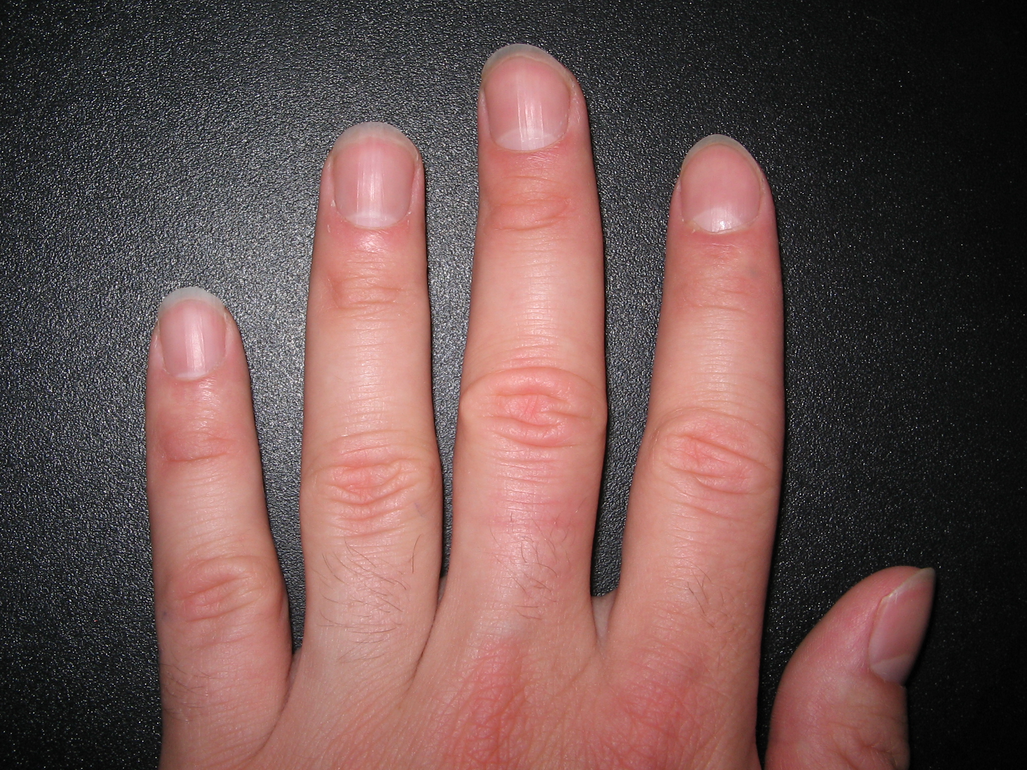
 Fingernails
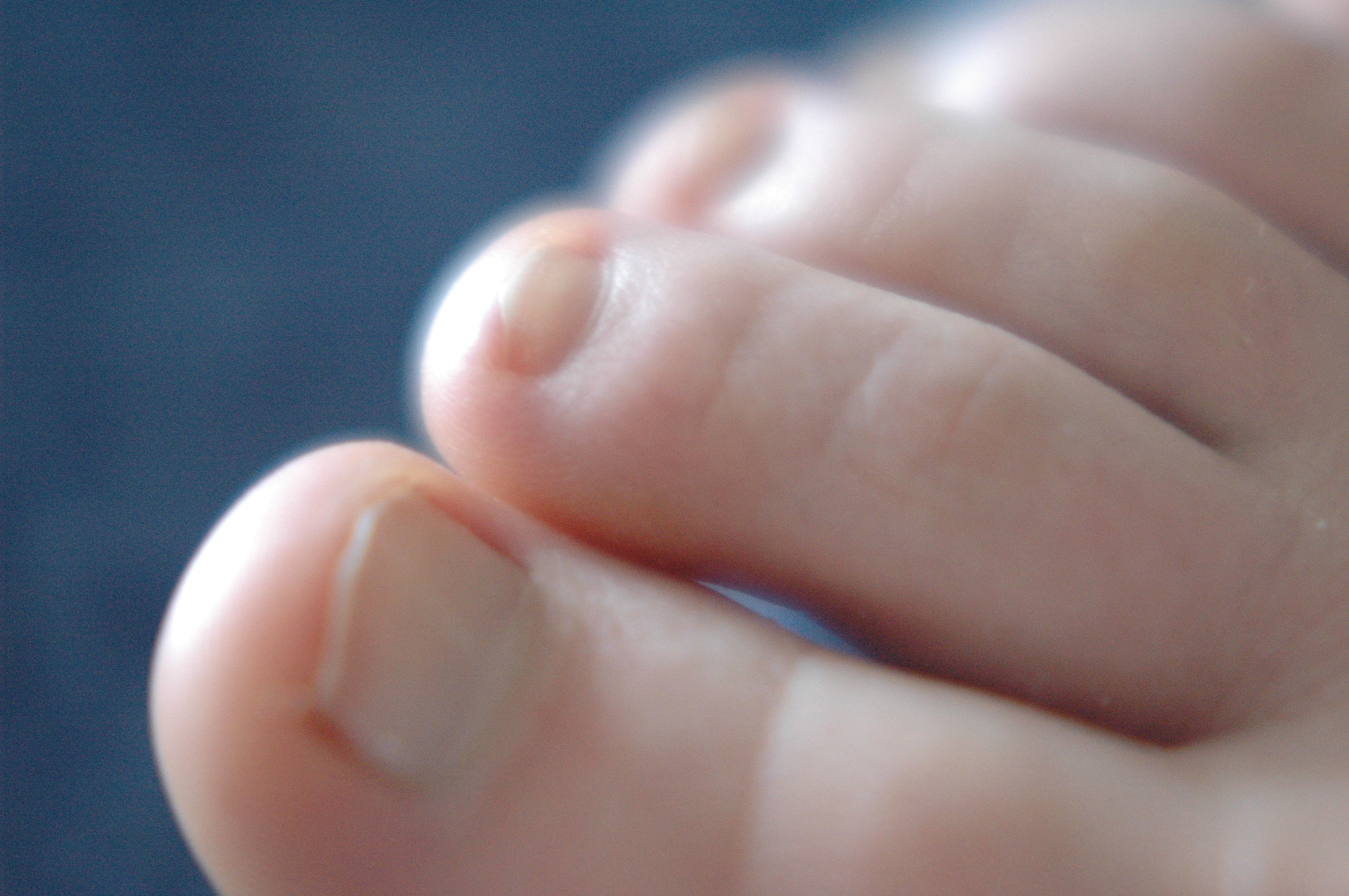
 Toenails
A nail is a horn-like envelope covering the dorsal aspect of the terminal phalanges of fingers and toes in humans, most non-human primates, and a few other mammals. Nails are similar to claws, which are found on numerous other animals. Fingernails and toenails are made of a tough protein called keratin, as are animals' hooves and horns. The mammalian nail, claw, and hoof are all examples of unguis [plural ungues].
Contents
Human anatomy
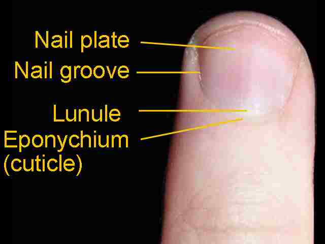
The nail consists of the nail plate, the nail matrix and the nail bed below it, and the grooves surrounding it.[1]
Parts of the nail
Nail 
Basic nail anatomy Latin unguis Code TH H3.12.00.3.02001 The matrix (synonyms:[2] matrix unguis, keratogenous membrane, nail matrix, onychostroma) is the tissue (or germinal matrix) upon which the nail rests,[3] the part of the nail bed that extends beneath the nail root and contains nerves, lymph and blood vessels.[4] The matrix is responsible for the production of the cells that become the nail plate. The width and thickness of the nail plate is determined by the size, length, and thickness of the matrix, while the shape of the fingertip itself determines if the nail plate is flat, arched or hooked.[5] The matrix will continue to grow as long as it receives nutrition and remains in a healthy condition.[4] As new nail plate cells are incubated, they emerge from the matrix round and white to push older nail plate cells forward; and in this way yet older cells become compressed, flat, and translucent, making the pink colour of the capillaries in the nail bed below visible.[6]
The lunula (occasionally called simply "the moon") is the visible part of the matrix, the whitish crescent-shaped base of the visible nail.[7] The lunula is largest in the thumb and often absent in the little finger.
The nail bed is the skin beneath the nail plate.[7] Like all skin, it is composed of two types of tissues: the deeper dermis, the living tissue fixed to the bone which contains capillaries and glands,[8] and the superficial epidermis, the layer just beneath the nail plate which moves forward with the plate. The epidermis is attached to the dermis by tiny longitudinal "grooves"[5] known as the matrix crests or crests of nail matrix (cristae matricis unguis).[3][8] During old age, the plate thins and these grooves are made evident in the structure.[5]
The nail sinus (sinus unguis) is the deep furrow into which the nail root is inserted.[3]
The nail root (radix unguis) is the part of nail situated in the nail sinus,[3] i.e. the base of the nail embedded underneath the skin. It originates from the actively growing tissue below, the matrix.[4]
The nail plate or body of nail (corpus unguis)[3] is the actual nail, and like hair and skin, made of translucent keratin protein made of amino acids. In the nail it forms a strong flexible material made of several layers of dead, flattened cells.[5] The plate appears pink because of the underlying capillaries.[7] Its (transversal) shape is determined by the form of the underlying bone.[5] In common usage, the word nail often refers to the this part only.
The free margin (margo liber) or distal edge is the anterior margin of the nail plate corresponding to the abrasive or cutting edge of the nail.[3] The hyponychium (informally known as the "quick")[9] is the epithelium located beneath the nail plate at the junction between the free edge and the skin of the fingertip. It forms a seal that protects the nail bed.[4] The onychodermal band is the seal between the nail plate and the hyponychium. It is found just under the free edge, in that portion of the nail where the nail bed ends and can be recognized by its glassy, greyish colour (in fair-skinned people). It is not perceptible in some individuals while it is highly prominent on others.[5]
The eponychium is the small band of epithelium that extends from the posterior nail wall onto the base of the nail.[3] Often and erroneously[contradictory] called the "proximal fold" or "cuticle", the eponychium is the end of the proximal fold that folds back upon itself to shed an epidermal layer of skin onto the newly formed nail plate. This layer of non-living, almost invisible skin is the cuticle that "rides out" on the surface of the nail plate. Together, the eponychium and the cuticle form a protective seal. The cuticle on the nail plate is dead cells and is often removed during manicure, but the eponychium is living cells and should not be touched.[6] The perionyx is the projecting edge of the eponychium covering the proximal strip of the lunula.[3]
The nail wall (vallum unguis) is the cutaneous fold overlapping the sides and proximal end of the nail. The lateral margin (margo lateralis) is lying beneath the nail wall on the sides of the nail and the nail groove or fold (sulcus matricis unguis) are the cutaneous slits into which the lateral margins are embedded.[3]
The paronychium is the border tissue around the nail[10] and paronychia is an infection in this area.
Function
A healthy (finger)nail has the function of protecting the distal phalanx, the fingertip, and the surrounding soft tissues from injuries. It also serves to enhance precise delicate movements of the distal digits through counter-pressure exerted on the pulp of the finger. [1] The nail then acts as a counterforce when the end of the finger touches an object, thereby enhancing the sensitivity of the fingertip,[11] even though there are no nerve endings in the nail itself. Finally, the nail functions as a tool, enabling for instance a so called "extended precision grip" (e.g. pulling out a splinter in one's finger).
Growth
The growing part of the nail is the part still under the skin at the nail's proximal end under the epidermis, which is the only living part of a nail.
In mammals, the length and growth rate of nails is related to the length of the terminal phalanges (outermost finger bones). Thus, in humans, the nail of the index finger grows faster than that of the little finger; and fingernails grow up to four times faster than toenails. [12]
In humans, nails grow at an average rate of 3 mm (0.12 in) a month (as they are a form of hair).[13] Fingernails require 3 to 6 months to regrow completely, and toenails require 12 to 18 months. Actual growth rate is dependent upon age, gender, season, exercise level, diet, and hereditary factors. Nails grow faster in the summer than in any other season.[14] Contrary to popular belief, nails do not continue to grow after death; the skin dehydrates and tightens, making the nails (and hair) appear to grow.[15]
Medical aspects
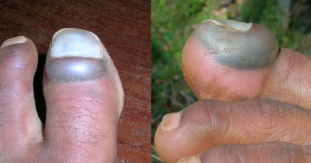
 Mechanical injury can result in the nail being dropped.
Healthcare and pre-hospital-care providers (EMTs or paramedics) often use the fingernail beds as a cursory indicator of distal tissue perfusion of individuals that may be dehydrated or in shock.[16] However, this test is not considered reliable in adults.[17] This is known as the CRT or blanch test.
WEJ Procedure: briefly depress the fingernail bed gently with a finger. This will briefly turn the nailbed white; the normal pink colour should be restored within a second or two. Delayed return to pink colour can be an indicator of certain shock states such as hypovolemia [18][19]
Nail growth record can show the history of recent health and physiological imbalances, and has been used as a diagnostic tool since ancient times.[20] Deep transverse grooves known as Beau's lines may form across the nails (not along the nail from cuticle to tip) and are usually a natural consequence of aging, though they may result from disease. Discoloration, thinning, thickening, brittleness, splitting, grooves, Mees' lines, small white spots, receded lunula, clubbing (convex), flatness, spooning (concave) can indicate illness in other areas of the body, nutrient deficiencies, drug reaction or poisoning, or merely local injury. Nails can also become thickened (onychogryphosis), loosened (onycholysis), infected with fungus (onychomycosis) or degenerate (onychodystrophy); for further information see nail diseases.
Health and care
Bluish or purple fingernail beds may be a symptom of peripheral cyanosis, which indicates oxygen deprivation.
Nails can dry out, just like skin. They can also peel, break, and be infected. Toe infections, for instance, can be caused or exacerbated by dirty socks, specific types of aggressive exercise, tight footwear, and walking unprotected in an unclean environment.[citation needed]
Nail tools used by different people may transmit infections. Nail files, "if ... used on different people, ... may spread nail fungi, staph bacteria or viruses," warns Ted Dischman, a spokesperson for the California Board of Barbering and Cosmetology.[1] In fact, over 100 bacterial skin infections in 2000 were traced to footbaths in nail salons.[citation needed] To avoid this, new improved contactless tools can be used, for example, gel and cream cuticle removers instead of cuticle scissors.
Many people also compulsively bite their nails.
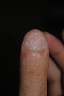 Damaged thumbnail
Nail disease can be very subtle and should be evaluated by a dermatologist with a focus in this particular area of medicine.[21] However, most times it is a nail technician who will note a subtle change in nail disease.
Inherited accessory nail of the fifth toe occurs where the toenail of the smallest toe is separated, forming a smaller, "sixth toenail" in the outer corner of the nail.[citation needed] Like any other nail, it can be cut using a nail clipper.
Nutrition for healthy nails
Vitamin A is an essential micronutrient for vision, reproduction, cell and tissue differentiation, and immune function. Vitamin D and calcium work together in cases of maintaining homeostasis, creating muscle contraction, transmission of nerve pulses, blood clotting, and membrane structure. A lack of vitamin A, vitamin D, and calcium can cause dryness and brittleness. Sources of these micronutrients include fortified milk, cereal, and juices, salt-water fish, fish-liver oils, and some vegetables. Vitamin B12 is mainly found in animal sources such as liver and kidney, fish, chicken, and dairy products and therefore can cause intake issues in vegan populations. Not enough B12 vitamin can lead to excessive dryness, darkened nails, and rounded or curved nail ends. Insufficient intake of both vitamin A and B, as previously described, results in fragile nails with horizontal and vertical ridges.[22] Protein is a building material for new nails, therefore low dietary protein intake may cause white nail beds. Dietary sources of this macronutrient include eggs, milk, cheese, meat, beans and legumes. A lack of protein combined with deficiencies in folic acid and vitamin C produce hangnails. Essential fatty acids play a large role in healthy skin as well as nails. As touched upon previously, essential fatty acids can be obtained through consumption of fish, flaxseed, canola oil, seeds, leafy vegetables, and nuts. Splitting and flaking of nails may be due to a lack of linoleic acid. Iron-deficiency anemia can lead to a pale color along with a thin, brittle, ridged texture. Iron deficiency in general may cause the nails to become flat or concave, rather than convex. Iron can be found in animal sources, called heme iron, such as meat, fish, and poultry, and can also be found in fruits, vegetables, dried beans, nuts, and grain products, also known as non-heme iron. Heme iron is absorbed fairly easily in comparison to non-heme iron, however both types provide the necessary bodily functions.[23]
Fashion
Manicures and pedicures are health and cosmetic procedures to groom, trim, and paint the nails and manage calluses. They require various tools such as cuticle scissors, nail scissors, nail clippers, and nail files. Artificial nails can also be appended onto real nails for cosmetic purposes.
A person whose occupation is to cut, shape and care for nails as well as to apply overlays such as acrylic and UV Gel is sometimes called a nail technician. The place where a nail technician works may be a nail salon or nail shop or nail bar.
Painting the nails with nail polish (also called nail lacquer and nail varnish) is a common practice dating back to at least 3000 B.C.
Non-human anatomy
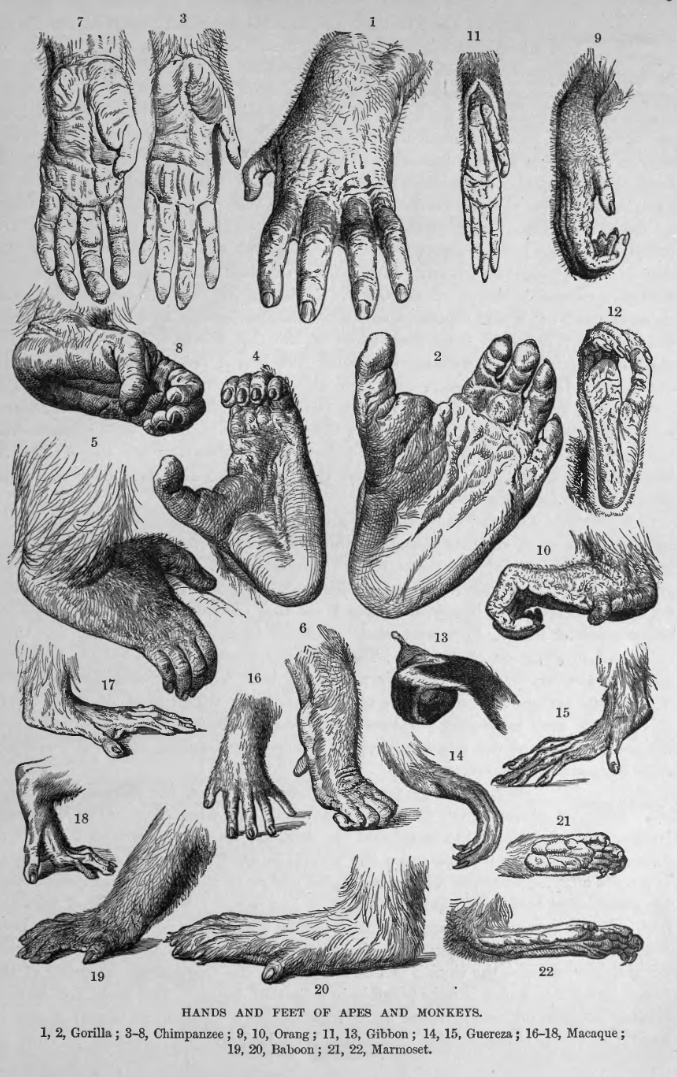
 Nails are a distinguishing feature in the primate order.
The nails of primates and the hooves of running mammals evolved from the claws of reptiles.[24]
In contrast to nails, claws are typically curved ventrally (downwards in animals) and compressed sideways. They serve a multitude of functions — including climbing, digging, and fighting — and have undergone numerous adaptive changes in different animal taxa. Claws are pointed at their ends and are composed of two layers: a thick, deep layer and a superficial, hardened layer which serves a protective function. The underlying bone is a virtual mould of the overlying horny structure and therefore has the same shape as the claw or nail. Compared to claws, nails are flat, less curved, and do not extend far beyond the tip of the digits. The ends of the nails usually consist only of the "superficial", hardened layer and are not pointed like claws.[24]
With only a few exceptions, primates retain plesiomorphic (original, "primitive") hands with five digits, each equipped with either a nail or a claw. For example, all prosimians (i.e. "primitive" primates or "proto-primates") have nails on all digits except the second toe which is equipped with a so called toilet-claw (i.e. important for grooming activities). The needle-clawed bushbaby (Euoticus) have keeled nails (the thumb and the first and the second toes have claws) featuring a central ridge that ends in a needle-like tip. In tree shrews all digits have claws and, unlike most primates, the digits of their feet are positioned close together, and therefore the thumb cannot be brought into opposition (another distinguishing feature of primates).[24]
A study of the fingertip morphology of four small-bodied New World monkey species indicated a correlation between increasing small-branch foraging and
- expanded apical pads,
- developed epidermal ridges (fingerprints),
- broadened distal parts of distal phalanges (fingertip bone), and
- reduced flexor and extensor tubercles.
This suggests that whereas claws are useful on large-diameter branches, wide fingertips with nails and epidermal ridges were required for habitual locomotion on small-diameter branches. It also indicates keel-shaped nails of Callitrichines (a family of New World monkeys) is a derived postural adaptation rather than retained ancestral condition. [25]
See also
- List of cutaneous conditions
- Nail fetish
References
- ^ a b Onumah, Neh; Scher, Richard K (May 2009). "Nail Surgery". eMedicine. http://emedicine.medscape.com/article/1126725-overview. Retrieved March 2010.
- ^ "Nail matrix". Biology Online. 2005. http://www.biology-online.org/dictionary/Nail_matrix. Retrieved February 2010.
- ^ a b c d e f g h i Feneis, Heinz (2000). Pocket Atlas of Human Anatomy (4th ed.). Thieme. pp. 392–95. ISBN 3-13-511204-7.
- ^ a b c d "Glossary of Nail Technology Terminology". 2008. https://www.nailsuperstore.com/tips/view.aspx?TipId=81. Retrieved February 2010.
- ^ a b c d e f Preuss, Marti (2000). "Understanding Your Natural Nails". Hooked on Nails. http://hooked-on-nails.com/naturalnails.html. Retrieved February 2010.
- ^ a b Lellipop (August 2006). "Anatomy of the nail". Salon Geek. http://www.salongeek.com/health-safety-unatural/40362-anatomy-nail.html. Retrieved February 2010.
- ^ a b c "Nail Anatomy". Nail Doctors. 2005. http://www.naildoctors.com/nail_anatomy.html. Retrieved February 2010.
- ^ a b "Glossary fo Nail Conditions". The Achilles Foot Health Centre. http://www.footdoc.ca/Website%20Nail%20Conditions%20(A%20Glossary).htm.
- ^ http://books.google.com/books?id=p8VqAAAAMAAJ&q=hyponychium+quick&dq=hyponychium+quick&hl=en&ei=0oSuTMSqA4O88gao4PTcBA&sa=X&oi=book_result&ct=result&resnum=1&ved=0CCUQ6AEwAA
- ^ Jordan, Christopher; Mirzabeigi, Edwin (2000-04-01). Atlas of orthopaedic surgical exposures. Thieme. p. 101. ISBN 0865777764. http://books.google.com/?id=gMSW59keA4UC&pg=PA101.
- ^ Wang, Quincy C; Johnson, Brett A (May 2001). "Fingertip Injuries". American Family Physician. http://www.aafp.org/afp/20010515/1961.html. Retrieved March 2010.
- ^ Ravosa, Matthew J.; Dagosto, Marian (2007). Primate origins: adaptations and evolution. Springer. pp. 389–90. ISBN 0387303359. http://books.google.com/?id=C8RAGvdzedoC&pg=PA416.
- ^ Toenail Definition - Medicine.net
- ^ Hunter, J. A. A., Savin, J., & Dahl, M. V. (2002). Clinical dermatology. Malden, Mass: Blackwell Science. p. 173. ISBN 0632059168
- ^ BMJ 2007;335(7633):1288 (22 December), doi:10.1136/bmj.39420.420370.25
- ^ Monterey County EMS Manual. Chapter XI, Patient assessment.
- ^ Schriger DL, Baraff LJ (Jun 1991). "Capillary refill—is it a useful predictor of hypovolemic states?". Ann Emerg Med 20 (6): 601–5. doi:10.1016/S0196-0644(05)82375-3. PMID 2039096. http://linkinghub.elsevier.com/retrieve/pii/S0196-0644(05)82375-3.
- ^ MedlinePlus Encyclopedia Capillary nail refill test
- ^ St. Luke's Hospital. Capillary nail refill test.
- ^ American Academy of Dermatology - Nail Health
- ^ http://www.nailsmag.com/feature.aspx?fid=762&ft=1
- ^ There are some over the counter vitamin supplements that may help in growth of strong nails such as certain Multivitamins and Biotin, though this is quite subjective as well. Zempleni, J; R.B. Rucker, D.B. McCormick, J.W. Suttie (2007). Handbook of vitamins (4th ed.). http://linus.lmu.edu.
- ^ Cashman MW, Sloan SB (2010). "Nutrition and nail disease". Clinics in Dermatology 28 (4): 420–425. doi:10.1016/j.clindermatol.2010.03.037. PMID 20620759.
- ^ a b c Ankel-Simons, Friderun (2007). Primate anatomy: an introduction (3rd ed.). pp. 342–44. ISBN 0-12-372576-3.
- ^ Hamrick, Mark W. (1998). "Functional and adaptive significance of primate pads and claws: Evidence from New World anthropoids". American Journal of Physical Anthropology (Wiley-Liss) 106 (2): 113–127. doi:10.1002/(SICI)1096-8644(199806)106:2<113::AID-AJPA2>3.0.CO;2-R. PMID 9637179. http://www3.interscience.wiley.com/journal/28218/abstract. (Abstract)
Integumentary system (TA A16, TH H3.12, GA 10.1062) Skin Basement membrane zoneSkin fieldsHeadcampus frontalis, campus parietalis, campus occipitalis, campus temroralis, campus facialis (campus orbitalis, campus nasalis, campus oralis, campus mentalis, campus infraorbitalis, campus buccalis, campus zygomaticus)Neckcampus cervicalis anterior (campus submandibularis, campus caroticus, campus omotrachealis, campus submentalis), campus sternocleidomastoideus, campus cervicalis posterior (campus omoclavicularis), campus nuchalisChestcampus presternalis, campus clavipectoralis, campus pectoralis verus, campus mammarius, campus inframammarius, campus axillarisAbdomencampus hypochondriacus, campus epigastricus, campus abdominalis lateralis, campus umbilicalis, campus inguinalis, campus hypogastricusPerineumcampus analis, campus urogenitalisSubcutaneous tissue Panniculus/Pannus (Panniculus adiposus · Panniculus carnosus) · Stratum membranosum · Loose connective tissue · Superficial fasciaAdnexa Skin glandsSweat glands: Apocrine sweat gland · Eccrine sweat gland
SebaceousHair shaftArrector pili musclePilosebaceous unitHair sebaceous glandNailExternal links
- Nail Disorders and Diseases - types of nail disorders: Paronychia, Onychogryphosis, Melanonychia
Categories:- Nails (anatomy)


Wikimedia Foundation. 2010.