- Basement membrane
-
Basement membrane 
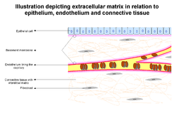
Illustration depicting basement membrane in relation to epithelium and endothelium. Also seen are other extracellular matrix components Latin membrana basalis Code TH H2.00.00.0.00005 The basement membrane is a thin sheet of fibers that underlies the epithelium, which lines the cavities and surfaces of organs including skin, or the endothelium, which lines the interior surface of blood vessels.
Contents
Composition
The basement membrane is the fusion of two lamina, the basal lamina and the reticular lamina (or lamina reticularis). The lamina reticularis is attached to the basal lamina with anchoring fibrils (type VII collagen fibers) and microfibrils (fibrillin). The two layers are collectively known as the basement membrane.[1]
The basal lamina layer can further be divided into two layers. The clear layer closer to the epithelium is called the lamina lucida, while the dense layer closer to the connective tissue is called the lamina densa. The electron-dense lamina densa membrane is about 30–70 nanometers in thickness, and consists of an underlying network of reticular collagen (type IV) fibrils (fibroblast precursors) which average 30 nanometers in diameter and 0.1–2 micrometers in thickness. In addition to collagen, this supportive matrix contains intrinsic macromolecular components.
The Lamina Densa (which is made up of type IV collagen fibers; perlecan (a heparan sulfate proteoglycan)[2] coats these fibers and they are high in heparan sulfate) and the Lamina Lucida (made up of laminin, integrins, entactins, and dystroglycans) together make up the basal lamina.
To represent the above in a visually organised manner, the basement membrane is organized as follows:
- Epithelial Tissue (outer)
- Basement Membrane
- Basal Lamina
- Lamina Lucida
- laminin
- integrins
- entactins
- dystroglycans
- Lamina Densa
- Type IV collagen (coated with perlecan, rich in heparan sulfate)
- Lamina Lucida
- Attaching proteins (between Basal and Reticular Laminae)
- Type VII collagen (anchoring fibrils)
- fibrillin (microfibrils)
- Lamina Reticularis
- ??
- Basal Lamina
- Connective Tissue (inner)
Function and importance
The primary function of the basement membrane is to anchor down the epithelium to its loose connective tissue underneath. This is achieved by cell-matrix adhesions through substrate adhesion molecules (SAMs).
The basement membrane acts as a mechanical barrier, preventing malignant cells from invading the deeper tissues.[3] Early stages of malignancy that are thus limited to the epithelial layer by the basement membrane are called carcinoma in situ.
The basement membrane is also essential for angiogenesis (development of new blood vessels). Basement membrane proteins have been found to accelerate differentiation of endothelial cells.[4]
The most notable examples of basement membranes is in the glomerular filtration of the kidney, by the fusion of the basal lamina from the endothelium of glomerular capillaries and the basal lamina of the epithelium of the Bowman's capsule, and between lung alveoli and pulmonary capillaries, by the fusion of the basal lamina of the lung alveoli and of the basal lamina of the lung capillaries, which is where oxygen and CO2 diffusion happens.
Diseases
Some diseases result from a poorly functioning basement membrane. The cause can be genetic defects, injuries by the body's own immune system, or other mechanisms.[5]
Genetic defects in the collagen fibers of the basement membrane cause Alport syndrome.
Non-collagenous domain basement membrane collagen type IV is autoantigen (target antigen) of autoantibodies in the autoimmune disease Goodpasture's syndrome.[6]
A group of diseases stemming from improper function of basement membrane zone are united under the name epidermolysis bullosa.
See also
- Intima
References
- ^ M Paulsson; Basement membrane proteins: structure, assembly, and cellular interactions; Critical Reviews in Biochemistry and Molecular Biology, Vol 27, Issue 1, 93-127, 1992
- ^ DM Noonan, A Fulle, P Valente, S Cai, E Horigan, M Sasaki, Y Yamada and JR Hassell; The complete sequence of perlecan, a basement membrane heparan sulfate proteoglycan, reveals extensive similarity with laminin A chain, low density lipoprotein-receptor, and the neural cell adhesion molecule; J. Biol. Chem., Vol. 266, Issue 34, 22939-22947, 12, 1991
- ^ L. A. Liotta, K. Tryggvason, S. Garbisa, Ian Hart, C. M. Foltz & S. Shafie; Metastatic potential correlates with enzymatic degradation of basement membrane collagen; Nature 284, 67 - 68 (6 March 1980).
- ^ Y Kubota, HK Kleinman, GR Martin and TJ Lawley; Role of laminin and basement membrane in the morphological differentiation of human endothelial cells into capillary-like structures; The Journal of Cell Biology, Vol 107, 1589-1598.
- ^ What’s Wrong With Summer Stiers? By ROBIN MARANTZ HENIG The New York Times, February 22, 2009
- ^ Janeway, Travers, Walport, Shlomchik; Immunobiology 5th ed.
Further reading
- Kefalides, Nicholas A. & Borel, Jacques P., ed (2005). Basement membranes: cell and molecular biology. Gulf Professional Publishing. ISBN 9780121533564. http://books.google.com/books?id=RM-FVY47NEgC.
Integumentary system (TA A16, TH H3.12, GA 10.1062) Skin Basement membrane zoneSkin fieldsHeadcampus frontalis, campus parietalis, campus occipitalis, campus temroralis, campus facialis (campus orbitalis, campus nasalis, campus oralis, campus mentalis, campus infraorbitalis, campus buccalis, campus zygomaticus)Neckcampus cervicalis anterior (campus submandibularis, campus caroticus, campus omotrachealis, campus submentalis), campus sternocleidomastoideus, campus cervicalis posterior (campus omoclavicularis), campus nuchalisChestcampus presternalis, campus clavipectoralis, campus pectoralis verus, campus mammarius, campus inframammarius, campus axillarisAbdomencampus hypochondriacus, campus epigastricus, campus abdominalis lateralis, campus umbilicalis, campus inguinalis, campus hypogastricusPerineumcampus analis, campus urogenitalisSubcutaneous tissue Panniculus/Pannus (Panniculus adiposus · Panniculus carnosus) · Stratum membranosum · Loose connective tissue · Superficial fasciaAdnexa Skin glandsSweat glands: Apocrine sweat gland · Eccrine sweat gland
SebaceousHair shaftArrector pili musclePilosebaceous unitHair sebaceous gland
This anatomy article is a stub. You can help Wikipedia by expanding it.
