- Lingual septum
-
Lingual septum 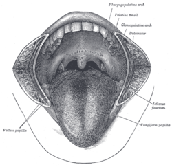
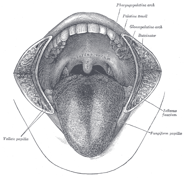
The mouth cavity. The cheeks have been slit transversely and the tongue pulled forward. (Lingual septum is visible at center of tongue, but not labeled.) 
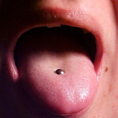
A pierced tongue, which has not accommodated for swelling Latin septum linguae Gray's subject #242 1132 The lingual septum consists of a vertical layer of fibrous tissue, extending throughout the entire length of the median plane of the tongue, though not quite reaching the dorsum.
It is thicker behind than in front, and occasionally contains a small fibrocartilage, about 6 mm. in length.
It is well displayed by making a vertical section across the organ.
See also
This article was originally based on an entry from a public domain edition of Gray's Anatomy. As such, some of the information contained within it may be outdated.
Parotid gland/Parotid duct · Submandibular gland/Submandibular duct · Sublingual gland/Major sublingual ductTeethsee tooth anatomydorsum (Taste bud, Median sulcus, Terminal sulcus, Foramen cecum, Lingual tonsils) · underside (Frenulum, Fimbriated fold, Sublingual caruncle) · Anterior · Posterior · Glossoepiglottic folds · Lingual septumOro-pharynx/
faucesOropharyngeal isthmus/Isthmus of the fauces
Soft palate (Uvula, Palatoglossal arch, Palatopharyngeal arch, Plica semilunaris of the fauces)
Palatine tonsilCategories:- Anatomy stubs
- Tongue
Wikimedia Foundation. 2010.