- Myocarditis
-
Myocarditis Classification and external resources
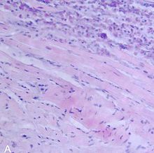
Histopathological image of myocarditis at autopsy in a patient with acute onset of congestive heart failure.ICD-10 I09.0, I51.4 ICD-9 391.2, 422, 429.0 DiseasesDB 8716 MedlinePlus 000149 eMedicine med/1569 emerg/326 MeSH D009205 Myocarditis is inflammation of heart muscle (myocardium). It resembles a heart attack but coronary arteries are not blocked.
Myocarditis is most often due to infection by common viruses, such as parvovirus B19, less commonly non-viral pathogens such as Borrelia burgdorferi (Lyme disease) or Trypanosoma cruzi, or as a hypersensitivity response to drugs.[1]
The definition of myocarditis varies, but the central feature is an infection of the heart, with an inflammatory infiltrate, and damage to the heart muscle, without the blockage of coronary arteries that define a heart attack (myocardial infarction) or other common non-infectious causes.[2] Myocarditis may or may not include death (necrosis) of heart tissue. It may include dilated cardiomyopathy.[1]
Myocarditis is often an autoimmune reaction. Streptococcal M protein and coxsackievirus B have regions (epitopes) that are immunologically similar to cardiac myosin. After the virus is gone, the immune system may attack cardiac myosin.[1]
Because a definitive diagnosis requires a heart biopsy, which doctors are reluctant to do because they are invasive, statistics on the incidence of myocarditis vary widely.[1]
The consequences of myocarditis thus also vary widely. It can cause a mild disease without any symptoms that resolves itself, or it may cause chest pain, heart failure, or sudden death. An acute myocardial infarction-like syndrome with normal coronary arteries has a good prognosis. Heart failure, even with dilated left ventricle, may have a good prognosis. Ventricular arrhythmias and high-degree heart block have a poor prognosis. Loss of right ventricular function is a strong predictor of death.[1]
Contents
Signs and symptoms
The signs and symptoms associated with myocarditis are varied, and relate either to the actual inflammation of the myocardium, or the weakness of the heart muscle that is secondary to the inflammation. Signs and symptoms of myocarditis include:[3]
- Chest pain (often described as "stabbing" in character)
- Congestive heart failure (leading to edema, breathlessness and hepatic congestion)
- Palpitations (due to arrhythmias)
- Sudden death (in young adults, myocarditis causes up to 20% of all cases of sudden death)[4]
- Fever (especially when infectious, e.g. in rheumatic fever)
- Symptoms in infants and toddlers tend to be more non-specific with generalized malaise, poor appetite, abdominal pain, chronic cough. Later stages of the illness will present with respiratory symptoms with increased work of breathing and is often mistaken for asthma.
Since myocarditis is often due to a viral illness, many patients give a history of symptoms consistent with a recent viral infection, including fever, rash, diarrhea, joint pains, and easy fatigueability.
Myocarditis is often associated with pericarditis, and many patients present with signs and symptoms that suggest concurrent myocarditis and pericarditis.
Causes
A large number of causes of myocarditis have been identified, but often a cause cannot be found. In Europe and North America, viruses are common culprits. Worldwide, however, the most common cause is Chagas' disease, an illness endemic to Central and South America that is due to infection by the protozoan Trypanosoma cruzi.[3]
Infections
- Viral (Parvovirus B19, Coxsackie virus, HIV, enterovirus, rubella virus, polio virus, cytomegalovirus, human herpesvirus 6 and possibly hepatitis C)
- Protozoan (Trypanosoma cruzi causing Chagas disease and Toxoplasma gondii)
- Bacterial (brucella, Corynebacterium diphtheriae, gonococcus, Haemophilus influenzae, Actinomyces, Tropheryma whipplei, Vibrio cholerae, Borrelia burgdorferi, leptospirosis, Rickettsia)
- Fungal (aspergillus)
- Parasitic (ascaris, Echinococcus granulosus, Paragonimus westermani, schistosoma, Taenia solium, Trichinella spiralis, visceral larva migrans, and Wuchereria bancrofti)
Bacterial myocarditis is rare in patients without immunodeficiency.
Toxins
- Drugs (ethanol, anthracyclines and some other forms of chemotherapy, and antipsychotics, e.g. clozapine, also some designer drugs such as mephedrone[5])
Immunologic
- Allergic (acetazolamide, amitriptyline)
- Rejection after a heart transplant
- Autoantigens (scleroderma, systemic lupus erythematosis, sarcoidosis, systemic vasculitis such as Churg-Strauss syndrome, Wegener's granulomatosis)
- Toxins (arsenic, toxic shock syndrome toxin, carbon monoxide, snake venom)
- Heavy metals (copper, iron)
Physical agents
Diagnosis
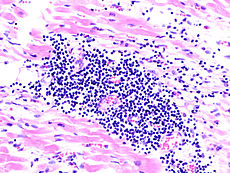
 Endomyocardial biopsy specimen. Extensive eosinophilic infiltrate involving the endocardium and myocardium (hematoxylin and eosin stain).
Myocarditis refers to an underlying process that causes inflammation and injury of the heart. It does not refer to inflammation of the heart as a consequence of some other insult. Many secondary causes, such as a heart attack, can lead to inflammation of the myocardium and therefore the diagnosis of myocarditis can not be made by evidence of inflammation of the myocardium alone.[6]
Myocardial inflammation can be suspected on the basis of electrocardiographic results (ECG), elevated C-reactive protein (CRP) and/or Erythrocyte sedimentation rate (ESR) and increased IgM (serology) against viruses known to affect the myocardium. Markers of myocardial damage (troponin or creatine kinase cardiac isoenzymes) are elevated.[3]
The electrocardiogram (ECG) findings most commonly seen in myocarditis are diffuse T wave inversions; saddle-shaped ST-segment elevations may be present (these are also seen in pericarditis).[3]
The gold standard is still biopsy of the myocardium, generally done in the setting of angiography. A small tissue sample of the endocardium and myocardium is taken, and investigated by a pathologist by light microscopy and—if necessary—immunochemistry and special staining methods. Histopathological features are: myocardial interstitium with abundant edema and inflammatory infiltrate, rich in lymphocytes and macrophages. Focal destruction of myocytes explains the myocardial pump failure.[3]
Cardiac magnetic resonance imaging (cMRI or CMR) has been shown to be very useful in diagnosing myocarditis by visualizing markers for inflammation of the myocardium.[7] Recently, consensus criteria for the diagnosis of myocarditis by CMR have been published [8]
Treatment
As most viral infections cannot be treated with directed therapy, symptomatic treatment is the only form of therapy for those forms of myocarditis.[9] In the acute phase, supportive therapy including bed rest is indicated. For symptomatic patients, digoxin and diuretics provide clinical improvement. For patients with moderate to severe dysfunction, cardiac function can be supported by use of inotropes such as Milrinone in acute phase followed by oral therapy with ACE inhibitors (Captopril, Lisinopril) when tolerated. People who do not respond to conventional therapy are candidates for bridge therapy with left ventricular assist devices (LVADs). Heart transplantation is reserved for patients who fail to improve with conventional therapy.[10]
In several small case series and randomized control trials, systemic corticosteroids have shown to have beneficial effects in patients with proven myocarditis.[10] However, data on the usefulness of corticosteroids should be interpreted with caution since 58% of adults recover spontaneously while most studies on children and infants lack control groups.[9]
Epidemiology
The exact incidence of myocarditis is unknown. However, in series of routine autopsies, 1–9% of all patients had evidence of myocardial inflammation. In young adults, up to 20% of all cases of sudden death are due to myocarditis.[3]
Among patients with HIV, myocarditis is the most common cardiac pathological finding at autopsy, with a prevalence of 50% or more.[1]
References
- ^ a b c d e f Leslie T. Cooper, Jr., (April 9, 2009) Myocarditis, N. Engl. J. Med. 360(15):1526-38
- ^ Kenneth L. Baughman, Special Report: Diagnosis of Myocarditis; Death of Dallas Criteria. Circulation. 2006;113:593-595 Free full text
- ^ a b c d e f Feldman AM, McNamara D (November 2000). "Myocarditis". N. Engl. J. Med. 343 (19): 1388–98. doi:10.1056/NEJM200011093431908. PMID 11070105.
- ^ Eckart RE, Scoville SL, Campbell CL, et al. (December 2004). "Sudden death in young adults: a 25-year review of autopsies in military recruits". Ann. Intern. Med. 141 (11): 829–34. PMID 15583223. http://www.annals.org/cgi/content/full/141/11/829.
- ^ Nicholson PJ, Quinn MJ, Dodd JD (December 2010). "Headshop heartache: acute mephedrone 'meow' myocarditis". Heart (British Cardiac Society) 96 (24): 2051–2. doi:10.1136/hrt.2010.209338. PMID 21062771.
- ^ Kumar, Vinay; Abbas, Abul K.; Fausto, Nelson; & Mitchell, Richard N. (2007). Robbins Basic Pathology (8th ed.). Saunders Elsevier. pp. 414-416 ISBN 978-1-4160-2973-1
- ^ Skouri HN, Dec GW, Friedrich MG, Cooper LT (2006). "Noninvasive imaging in myocarditis". J. Am. Coll. Cardiol. 48 (10): 2085–93. doi:10.1016/j.jacc.2006.08.017. PMID 17112998.
- ^ Friedrich MG, Sechtem U, Schulz-Menger J, Holmvang G, Alakija P, Cooper LT, White JA, Abdel-Aty H, Gutberlet M, Prasad S, Aletras AH, Laissy JP, Paterson I, Filipchuk NG, Kumar A, Pauschinger M, Liu P (2009). "Cardiovascular Magnetic Resonance in Myocarditis: A JACC White Paper". J. Am. Coll. Cardiol. 53 (17): 1475–87. doi:10.1016/j.jacc.2009.02.007. PMC 2743893. PMID 7351097. http://www.pubmedcentral.nih.gov/articlerender.fcgi?tool=pmcentrez&artid=2743893.
- ^ a b Hia, CP; Yip, WC, Tai, BC, Quek, SC (2004 Jun). "Immunosuppressive therapy in acute myocarditis: an 18 year systematic review". Archives of disease in childhood 89 (6): 580–4. doi:10.1136/adc.2003.034686. PMC 1719952. PMID 15155409. http://www.pubmedcentral.nih.gov/articlerender.fcgi?tool=pmcentrez&artid=1719952.
- ^ a b Aziz, KU; Patel, N, Sadullah, T, Tasneem, H, Thawerani, H, Talpur, S (2010 Oct). "Acute viral myocarditis: role of immunosuppression: a prospective randomised study". Cardiology in the young 20 (5): 509–15. doi:10.1017/S1047951110000594. PMID 20584348.
External links
Inflammation Acute preformed: Lysosome granules · vasoactive amines (Histamine, Serotonin)
synthesized on demand: cytokines (IFN-γ, IL-8, TNF-α, IL-1) · eicosanoids (Leukotriene B4, Prostaglandins) · Nitric oxide · KininsChronic Processes Traditional: Rubor · Calor · Tumor · Dolor (pain) · Functio laesa
Modern: Acute-phase reaction/Fever · Vasodilation · Increased vascular permeability · Exudate · Leukocyte extravasation · ChemotaxisSpecific locations CNS (Encephalitis, Myelitis) · Meningitis (Arachnoiditis) · PNS (Neuritis) · eye (Dacryoadenitis, Scleritis, Keratitis, Choroiditis, Retinitis, Chorioretinitis, Blepharitis, Conjunctivitis, Iritis, Uveitis) · ear (Otitis, Labyrinthitis, Mastoiditis)CardiovascularCarditis (Endocarditis, Myocarditis, Pericarditis) · Vasculitis (Arteritis, Phlebitis, Capillaritis)upper (Sinusitis, Rhinitis, Pharyngitis, Laryngitis) · lower (Tracheitis, Bronchitis, Bronchiolitis, Pneumonitis, Pleuritis) · MediastinitisDigestivemouth (Stomatitis, Gingivitis, Gingivostomatitis, Glossitis, Tonsillitis, Sialadenitis/Parotitis, Cheilitis, Pulpitis, Gnathitis) · tract (Esophagitis, Gastritis, Gastroenteritis, Enteritis, Colitis, Enterocolitis, Duodenitis, Ileitis, Caecitis, Appendicitis, Proctitis) · accessory (Hepatitis, Cholangitis, Cholecystitis, Pancreatitis) · PeritonitisArthritis · Dermatomyositis · soft tissue (Myositis, Synovitis/Tenosynovitis, Bursitis, Enthesitis, Fasciitis, Capsulitis, Epicondylitis, Tendinitis, Panniculitis)
Osteochondritis: Osteitis (Spondylitis, Periostitis) · Chondritisfemale: Oophoritis · Salpingitis · Endometritis · Parametritis · Cervicitis · Vaginitis · Vulvitis · Mastitis
male: Orchitis · Epididymitis · Prostatitis · Balanitis · Balanoposthitis
pregnancy/newborn: Chorioamnionitis · OmphalitisCategories:- Heart diseases
- Chronic rheumatic heart diseases
- Inflammations
Wikimedia Foundation. 2010.

