- Right ventricle
Infobox Anatomy
Name = PAGENAME
Latin = ventriculus dexter
GraySubject = 138
GrayPage = 531
.svg)
Caption = Anterior (frontal) view of the opened heart. White arrows indicate normal blood flow.
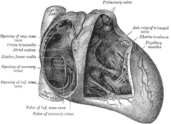
Caption2 = Interior of right side of heart.
Artery =right marginal branch of right coronary artery | System =
Precursor =primitive ventricle ,bulbus cordis
MeshName = Heart+Ventricles
MeshNumber = A07.541.560
The right ventricle is one of four chambers (two atria and two ventricles) in thehuman heart . It receivesdeoxygen atedblood from theright atrium via thecuspid valve , and pumps it into thepulmonary artery via thepulmonary valve andpulmonary trunk .It is triangular in form, and extends from the right atrium to near the
apex of the heart .Boundaries
Its anterosuperior surface is rounded and convex, and forms the larger part of the sternocostal surface of the heart.
Its under surface is flattened, rests upon the diaphragm, and forms a small part of the diaphragmatic surface of the heart.
Its posterior wall is formed by the
ventricular septum , which bulges into the right ventricle, so that a transverse section of the cavity presents a semilunar outline.Its upper and left angle forms a conical pouch, the
conus arteriosus , from which thepulmonary artery arises.A tendinous band, which may be named the tendon of the conus arteriosus, extends upward from the right atrioventricular fibrous ring and connects the posterior surface of the conus arteriosus to the aorta.
The wall of the right ventricle is thinner than that of the left, the proportion between them being as 1 to 3; it is thickest at the base, and gradually becomes thinner toward the apex.
The cavity equals in size that of the left ventricle, and is capable of containing about 85 c.c.
=Additionalee also
*
Left ventricle
*Double outlet right ventricle
*Right ventricular hypertrophy External links
*
Wikimedia Foundation. 2010.