- Neurulation
-
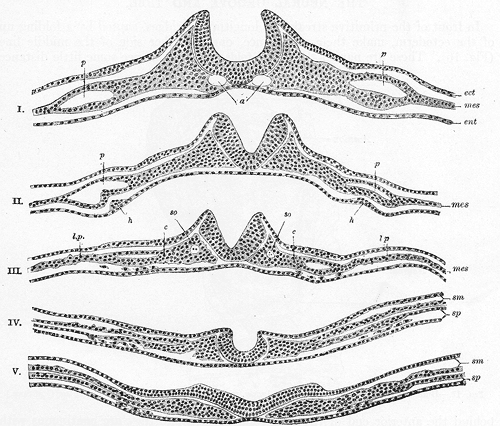
 Transverse sections that show the progression of the neural plate to the neural groove from bottom to top
Neurulation is the stage of organogenesis in vertebrate embryos, during which the neural tube is transformed into the primitive structures that will later develop into the central nervous system.[1]
The process begins when the notochord induces the formation of the central nervous system (CNS) by signaling the ectoderm germ layer above it to form the thick and flat neural plate. The neural plate folds in upon itself to form the neural tube, which will later differentiate into the spinal cord and the brain, eventually forming the central nervous system.[citation needed]
Different portions of the neural tube form by two different processes, called primary and secondary neurulation, in different species.[citation needed]
- In primary neurulation, the neural plate creases inward until the edges come in contact and fuse.
- In secondary neurulation, the tube forms by hollowing out of the interior of a solid precursor.
Contents
Primary neurulation
Induction
Primary neurulation occurs in response to soluble growth factors secreted by the notochord. Ectodermal cells are induced to form neuroectoderm from a variety of signals. Ectoderm sends and receives signals of BMP4 (bone morphogenic protein) and cells which receive BMP4 signal develop into epidermis. The inhibitory signals chordin, noggin and follistatin are needed to form neural plate. These inhibitory signals are created and emitted by the spemann organiser. Cells which do not receive BMP4 signaling due to the effects of the inhibitory signals will develop into the anterior neuroectoderm cells of the neural plate. Cells which receive FGF (fibroblast growth factor) in addition to the inhibitory signals form posterior neural plate cells.[citation needed]
Shape change
The cells of the neural plate are signaled to become high-columnar and can be identified through microscopy as different from the surrounding epiblastic ectoderm. The cells move laterally and away from the central axis and change into a truncated pyramid shape. This pyramid shape is achieved through tubulin and actin in the apical portion of the cell which constricts as they move. The variation in cell shapes is partially determined by the location of the nucleus within the cell, causing bulging in areas of the cells forcing the height and shape of the cell to change. This process is known as apical constriction[citation needed]
Folding
The process of the flat neural plate folding into the cylindrical neural tube is termed primary neurulation. As a result of the cellular shape changes, the neural plate forms the medial hinge point . The expanding epidermis puts pressure on the MHP and causes the neural plate to fold resulting in neural folds and the creation of the neural groove. The neural folds form dorsolateral hinge points (DLHP) and pressure on this hinge causes the neural folds to meet and fuse at the midline. The fusion requires the regulation of cell adhesion molecules. The neural plate switches from E-cadherin expression to N-cadherin and N-CAM expression to recognize each other as the same tissue and close the tube. This change in expression stops the binding of the neural tube to the epidermis. Neural plate folding is a complicated step.[citation needed]
The notochord plays an integral role in the development of the neural tube. Prior to neurulation, during the migration of epiblastic endoderm cells towards the hypoblastic endoderm, the notochordal process opens into an arch termed the notochordal plate and attaches overlying neuroepithelium of the neural plate. The notochordal plate then serves as an anchor for the neural plate and pushes the two edges of the plate upwards while keeping the middle section anchored. Some of the notochodral cells become incorporated into the center section neural plate to later form the floor plate of the neural tube. The notochord plate separates and forms the solid notochord.[citation needed]
The folding of the neural tube to form an actual tube does not occur all at once. Instead, it begins approximately at the level of the fourth somite at Carnegie stage 9 (around Embryonic day 20 in humans). The lateral edges of the neural plate touch in the midline and join together. This continues both cranially (toward the head) and caudally (toward the tail). The openings that are formed at the cranial and caudal regions are termed the cranial and caudal neuropores. In human embryos, the cranial neuropore closes approximately on day 24 and the caudal neuropore on day 26 (Carnegie stages 11 and 13 respectively). Failure of the cranial (anterior) and caudal (posterior) neuropore closure results in conditions called anecephaly and spina bifida, respectively. Additionally, failure of the neural tube to close throughout the length of the body results in a condition called cranioarchischisis.[citation needed]
Patterning
 Transverse section of the neural tube showing the floor plate and roof plate
After SHH from the notochord induces its formation, the floor plate of the incipient neural tube also secretes SHH. After closure, the neural tube forms a basal plate or floor plate and an alar plate or roof plate in response to the combined effects of Shh and factors including BMP4 secreted by the roof plate. The basal plate forms most of the ventral portion of the nervous system, including the motor portion of the spinal cord and brain stem; the alar plate forms the dorsal portions, devoted mostly to sensory processing.[citation needed]
The dorsal epidermis expresses BMP4 and BMP7. The roof plate of the neural tube responds to those signals to express more BMP4 and other TGF-b signals to form a dorsal/ventral gradient among the neural tube. The notochord expresses Sonic Hedgehog (Shh). The floor plate responds to Shh by producing its own Shh and forming a gradient. These gradients allows for the differential expression of transcription factors.[citation needed]
Complexities of the model
In actuality, the folding of the neural tube is still not entirely understood and is still being studied. The simplistic model of the closure occurring in one step cranially and caudally does not explain the high frequency of neural tube defects. Proposed theories include closure of the neural tube occurs in regions, rather than entirely linearly.[citation needed]
Secondary neurulation
In secondary neurulation, the neural ectoderm and some cells from the endoderm form the medullary cord. The medullary cord condenses, separates and then forms cavities. These cavities then merge to form a single tube. Secondary Neurulation occurs in the posterior section of most animals but it is better expressed in birds. Tubes from both primary and secondary neurulation eventually connect.[citation needed]
Early brain development
The anterior segment of the neural tube forms the three main parts of the brain: the forebrain, midbrain, and the hindbrain. Formation of these structures begins with a swelling of the neural tube in a pattern specified by Hox genes. Ion pumps are used to increase the fluid pressure within the tube and create a bulge. A blockage between the brain and the spinal cord prevents the fluid accumulation from leaking out. These brain regions further divide into subregions. The hindbrain divides into different segments called rhombomeres. Neural crest cells form ganglia above each rhombomere. The neural tube becomes the germinal neuroepithelium and serves as a source of new neurons during brain development. The brain develops from the inside-out.[citation needed]
Non-neural ectoderm tissue
Mesoderm surrounding the notochord at the sides will develop into the somites (future muscles, bones, and contributes to the formation of limbs of the vertebrate).[citation needed]
Neural crest cells
Masses of tissue called the neural crest that are located at the very edges of the lateral plates of the folding neural tube separate from the neural tube and migrate to become a variety of different but important cells.[citation needed]
Neural crest cells will migrate through the embryo and will give rise to several cell populations, including pigment cells and the cells of the peripheral nervous system.[citation needed]
Neural tube defects
Failure to complete the neurulation process will lead to an open neural tube, which is a common form of birth defect known as spina bifida.[2] Spina bifida can lead to paralysis beneath the affected region of the spinal cord. Sufferers may require crutches or wheelchairs to move about, and may also suffer from lack of bladder and bowel control.[citation needed]
Neural tube defects are among the most common and disabling birth defects, occurring in roughly 1 in every 500 live births.[3]
References
- ^ Slonim, Anthony D. & Pollack, Murray M., ed (2006). Pediatric critical care medicine. Lippincott Williams & Wilkins. p. 320. ISBN 9780781794695. http://books.google.com/books?id=_XavFllbnS0C&pg=PA320.
- ^ Squire, Larry R. (2003). Fundamental neuroscience. Academic Press. p. 370. ISBN 9780126603033. http://books.google.com/books?id=ZgUeT-SuR4sC&pg=PA370.
- ^ Daley, Darrel. Formation of the Nervous System. Last accessed on Oct 29, 2007.
Further reading
- Almeida, Karla L. et al. (2010). "Neural Induction". In Henning, Ulrich. Perspectives of Stem Cells: From Tools for Studying Mechanisms of Neuronal Differentiation Towards Therapy. Springer. ISBN 9789048133741. http://books.google.com/books?id=c0mFI5W2un0C&pg=PA1.
- Basch, Martín L. & Bonner-Fraser, Marianne (2006). "Neural Crest Inducing Signals". In Saint-Jennet, Jean-Pierre. Neural crest induction and differentiation. Springer. ISBN 9780387351360. http://books.google.com/books?id=KzF1LEEAnvAC&pg=PA24.
- Harland, Richard M. (1997). "Neural induction in Xenopus". In Cowan, W. Maxwell. Molecular and cellular approaches to neural development. Oxford University Press. ISBN 9780195111668. http://books.google.com/books?id=ZChr2WMbqzcC&pg=PA1.
- Ladher, Raj & Schoenwolf, Gary C. (2004). "Making a neural tube". In Jacobson, Marcus & Rao, Mahendra S.. Developmental neurobiology. Springer. ISBN 9780306483301. http://books.google.com/books?id=CcaSO-WJavIC&pg=PA1.
- Tian, Jing & Sampath, Karuna (2004). "Formation and Functions of the Floor Plate". In Gong, Zhiyuan & Korzh, Vladimir. Fish development and genetics: the zebrafish and medaka models. World Scientific. pp. 123; 139–140. ISBN 9789812388216.
- Zhang, Su-Chun (2005). "Neural specification from human embryonic stem cells". In Odorico, John S. et al. Human embryonic stem cells. Garland Science. ISBN 9781859962787. http://books.google.com/books?id=35VprwZsSRcC&pg=PA149.
External links
Developmental biology > Human embryogenesis (development of embryo) and development of fetus (TE E2.0) First three
weeksWeek 1Fertilization · Oocyte activation · Zygote · Cleavage · Morula · Blastula (Blastomere) · Blastocyst · Inner cell massWeek 2
(Bilaminar)Week 3
(Trilaminar)Archenteron/Primitive streak (Primitive pit, Primitive knot/Blastopore, Primitive groove) · Gastrula/Gastrulation · Regional specification · Embryonic discSplanchnopleuric mesenchymeChorda- · Paraxial (Somite/Somitomere) · Intermediate · Lateral plate (Intraembryonic coelom, Splanchnopleuric mesenchyme/Somatopleuric mesenchyme)Prenatal development/Mammalian development of nervous system (GA 9.733 and GA 10.1002, TE E5.13-16) Neurogenesis Rostral neuropore
Cephalic flexure · Pontine flexure
Alar plate (sensory) · Basal plate (motor)
Germinal matrixEye development Auditory development M: EYE
anat(g/a/p)/phys/devp/prot
noco/cong/tumr, epon
proc, drug(S1A/1E/1F/1L)
M: EAR
anat(e/p)/phys/devp
noco/cong, epon
proc, drug(S2)
Categories:- Neurulation
- Embryology of nervous system

Wikimedia Foundation. 2010.