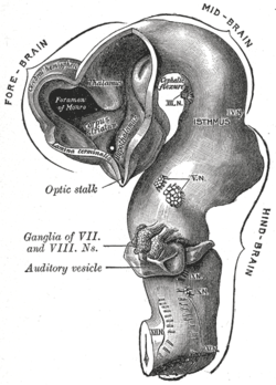
- Cephalic flexure
-
Cephalic flexure Brain of human embryo of four and a half weeks, showing interior of fore-brain. (Cephalic flexure visible at center top.) Latin flexura mesencephalica Gray's subject #184 737 Code TE E5.14.3.3.0.0.3 The mesencephalic flexure or cephalic flexure is the first flexure, or bend, of the embryonic brain; it appears in the region of the mid-brain. In human embryos it generally occurs at the end of the 3rd week/beginning of 4th.
By means of it the fore-brain is bent in a ventral direction around the anterior end of the notochord and fore-gut, with the result that the floor of the fore-brain comes to lie almost parallel with that of the hind-brain.
This flexure causes the mid-brain to become, for a time, the most prominent part of the brain, since its dorsal surface corresponds with the convexity of the curve. As a consequence, the cardiac plate, which was previously anterior to the forebrain, approximates its adult location in what will become the thoracic region.
External links
- Embryology at UNSW wwwpig/pigg/G7L
- Overview at nlm.nih.gov - online book
- Diagram at nlm.nih.gov - online book
This article was originally based on an entry from a public domain edition of Gray's Anatomy. As such, some of the information contained within it may be outdated.
Prenatal development/Mammalian development of nervous system (GA 9.733 and GA 10.1002, TE E5.13-16) Neurogenesis Cranial neural crest (Cardiac neural crest complex) · Truncal neural crestRostral neuropore
Cephalic flexure · Pontine flexure
Alar plate (sensory) · Basal plate (motor)
Germinal matrixEye development Auditory development M: EYE
anat(g/a/p)/phys/devp/prot
noco/cong/tumr, epon
proc, drug(S1A/1E/1F/1L)
M: EAR
anat(e/p)/phys/devp
noco/cong, epon
proc, drug(S2)
Categories:- Developmental biology stubs
- Neuroanatomy stubs
- Neuroanatomy
- Embryology of nervous system
Wikimedia Foundation. 2010.

