- Corneal epithelium
-
Corneal epithelium 
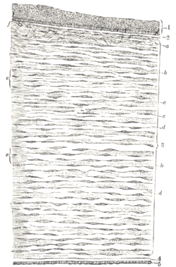
Vertical section of human cornea from near the margin. (Waldeyer.) Magnified.
1. Epithelium.
2. Anterior elastic lamina.
3. substantia propria.
4. Posterior elastic lamina.
5. Endothelium of the anterior chamber.
a. Oblique fibers in the anterior layer of the substantia propria.
b. Lamellæ the fibers of which are cut across, producing a dotted appearance.
c. Corneal corpuscles appearing fusiform in section.
d. Lamellæ the fibers of which are cut longitudinally.
e. Transition to the sclera, with more distinct fibrillation, and surmounted by a thicker epithelium.
f. Small blood vessels cut across near the margin of the cornea.Latin e. anterius corneae Gray's subject #225 1007 MeSH Epithelium,+corneal The corneal epithelium (epithelium corneæ anterior layer) is made up of epithelial tissue and covers the front of the cornea. It acts as a barrier to protect the cornea, resisting the free flow of fluids from the tears, and prevents bacteria from entering the epithelium and corneal stroma.
The corneal epithelium consists of several layers of cells. The cells of the deepest layer are columnar, known as basal cells. Then follow two or three layers of polyhedral cells, commonly known as wing cells. The majority of these are prickle cells, similar to those found in the stratum mucosum of the cuticle. Lastly, there are three or four layers of squamous cells, with flattened nuclei. The layers of the epithelium are constantly undergoing mitosis. Basal and wing cells migrate to the anterior of the cornea, while squamous cells age and slough off into the tear film.
Contents
Cornea Cell LASIK Complication
Epithelial ingrowth is a LASIK complication in which cells from the cornea surface layer (epithelial cells) begin to grow underneath the corneal flap. Epithelial ingrowth is a rarely occurring LASIK complication, appearing in less than one percent of LASIK procedures.[citation needed] However, the incidence of epithelial ingrowth appears to be higher after subsequent enhancement LASIK procedures.[citation needed] This complication is not present in PRK or other non-flap vision correction procedures.
See also
- Stratified squamous epithelium
Disorders
External links
Sensory system – visual system – globe of eye (TA A15.2.1–6, TH 3.11.08.0-5, GA 10.1005) Fibrous tunic (outer) Episcleral layer • Schlemm's canal • Trabecular meshworkUvea/vascular tunic (middle) Retina (inner) LayersCellsPhotoreceptor cells (Cone cell, Rod cell) → (Horizontal cell) → Bipolar cell → (Amacrine cell) → Retina ganglion cell (Midget cell, Parasol cell, Bistratified cell, Giant retina ganglion cells, Photosensitive ganglion cell) → Diencephalon: P cell, M cell, K cell
Muller gliaOtherAnterior segment Posterior segment Other M: EYE
anat(g/a/p)/phys/devp/prot
noco/cong/tumr, epon
proc, drug(S1A/1E/1F/1L)
Epithelial cells Surface epithelium Simple squamous epithelium (Endothelium, Mesothelium)
Pseudostratified columnar epithelium (Respiratory epithelium)
Stratified squamous epithelium
Stratified cuboidal epithelium
Stratified columnar epithelium
Transitional epithelium/UrotheliumGland/
glandular epitheliumMechanismShapeSecretionSerous glands · Mucous glandsComponentssee also Template:Epithelial neoplasms Categories:- Eye anatomy
Wikimedia Foundation. 2010.


