- Bowman's membrane
Infobox Anatomy
Name = Bowman's membrane
Latin = l. limitans anterior corneae
GraySubject = 225
GrayPage = 1008
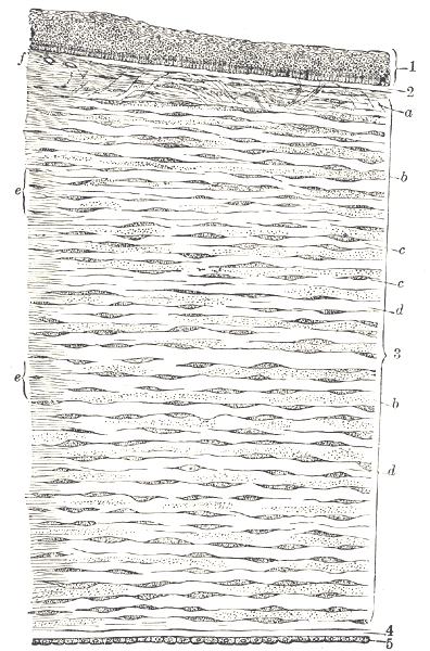
Caption = Vertical section of human cornea from near the margin. (Waldeyer.) Magnified.
1.Epithelium .
2. Anterior elastic lamina.
3.substantia propria .
4. Posterior elastic lamina.
5.Endothelium of theanterior chamber .
a. Oblique fibers in the anterior layer of thesubstantia propria .
b. Lamellæ the fibers of which are cut across, producing a dotted appearance.
c.Corneal corpuscles appearingfusiform in section.
d. Lamellæ the fibers of which are cut longitudinally.
e. Transition to thesclera , with more distinct fibrillation, and surmounted by a thickerepithelium .
f. Small bloodvessels cut across near the margin of thecornea .
Caption2 =
System =
Precursor =
MeshName = Bowman+membrane
MeshNumber = A09.371.060.217.113
DorlandsPre = l_02
DorlandsSuf = 12476272
The Bowman's membrane (Bowman's layer, anterior limiting lamina, anterior elastic lamina) is a smooth layer in theeye . It is located between the frontepithelium and the stroma in thecornea . It is composed of strongcollagen fibers and helps the cornea maintain its shape. If the Bowman's membrane is damaged, scarring would normally occur.In adult humans, this layer is 8-12 μm thick. [Hogan MJ, Alvarado JA, Weddell E: Histology of the Human Eye. Philadelphia: WB Saunders, 1971]
Bowman's layer is absent in cats, dogs, and other carnivores.Merindano MD; Costa J; Canals M; Potau JM, and Ruano D. [http://www.med.ub.es/sae/EJA/EJA_V6_N3_02.pdf#search=%22%22mammalian%20cornea%22%2C%20layers%22 "A comparative study of Bowman's layer in some mammals: Relationships with other constituent corneal structures."] "European Journal of Anatomy." Volume 6, Number 3, December 2002.]
The Bowman's membrane is named after Sir
William Bowman (1816 -1892 ), an Englishphysician ,anatomist andophthalmologist , who discovered this membrane.ee also
*
Refractive surgery
*Descemet's membrane References
External links
*
* [http://www.sheinman.com/Aanatomyp4.htm Diagram at sheinman.com]
* [http://www.laservue.net/images/cornea_crosssection_en.jpgDiagram at cornea_crosssection_en.jpg]
Wikimedia Foundation. 2010.