- Inner limiting membrane
-
Inner limiting membrane [[Image: |250px|]]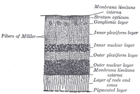

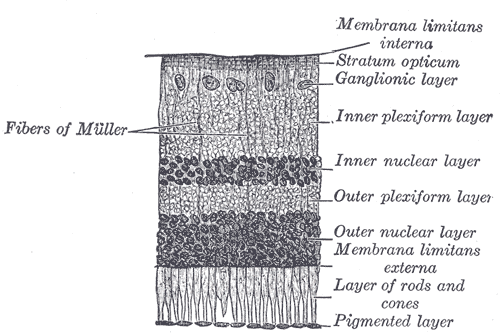
Section of retina. (Membrana limitans interna labeled at right, at top.) [[Image: |250px|]]
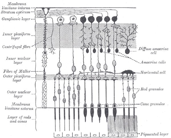
Plan of retinal neurons. (Membrana limitans interna labeled at left, at top.) Latin membrana limitans interna Gray's subject #225 1017 The inner limiting membrane is the boundary between the retina and the vitreous body, formed by astrocytes and the end feet of Müller cells. It is separated from the vitreous humor by a basal lamina.
External links
Sensory system – visual system – globe of eye (TA A15.2.1–6, TH 3.11.08.0-5, GA 10.1005) Fibrous tunic (outer) 

Uvea/vascular tunic (middle) Retina (inner) LayersCellsPhotoreceptor cells (Cone cell, Rod cell) → (Horizontal cell) → Bipolar cell → (Amacrine cell) → Retina ganglion cell (Midget cell, Parasol cell, Bistratified cell, Giant retina ganglion cells, Photosensitive ganglion cell) → Diencephalon: P cell, M cell, K cell
Muller gliaOtherAnterior segment Posterior segment Other M: EYE
anat(g/a/p)/phys/devp/prot
noco/cong/tumr, epon
proc, drug(S1A/1E/1F/1L)
Categories:- Eye anatomy
- Eye stubs

Wikimedia Foundation. 2010.