Midget cell

Midget cell

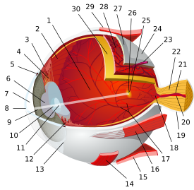
A midget cell is one type of retinal ganglion cell. Midget cells originate in the ganglion cell layer of the retina, and project to the parvocellular layers of the lateral geniculate nucleus (LGN). The axons of midget cells travel through the optic nerve and optic tract, ultimately synapsing with parvocellular cells in the LGN. These cells are known as midget retinal ganglion cells due to the small sizes of their dendritic trees and cell bodies. About 80% of RGCs are midget cells. They receive inputs from relatively few rods and cones. In many cases, they are connected to midget bipolar cells, which are linked to one cone each.[1] They have slow conduction velocity, and respond to changes in color but respond only weakly to changes in contrast unless the change is great.[2] They have simple center-surround receptive fields, where the center may be either ON or OFF while the surround is the opposite.

See also

References

  1. ^ "eye, human."Encyclopædia Britannica. 2008. Encyclopædia Britannica 2006 Ultimate Reference Suite DVD
  2. ^ Principles of Neural Science 4th Ed. Kandel et al.

Wikimedia Foundation. 2010.

Игры ⚽ Поможем написать реферат

Look at other dictionaries:

  • Cell — The basic structural and functional unit in people and all living things. Each cell is a small container of chemicals and water wrapped in a membrane. Each cell in the human body there are 100 trillion cells in each of us contains the entire… …   Medical dictionary

  • Photosensitive ganglion cell — Section of retina: light strikes first the ganglion cell layer, last the rods and cones Photosensitive ganglion cells, also called photosensitive Retinal Ganglion Cells (pRGC), intrinsically photosensitive Retinal Ganglion Cells (ipRGC) or… …   Wikipedia

  • Photoreceptor cell — This article is about cellular photoreceptors. For other types of photoreceptors, see Photoreceptor (disambiguation). Rods and cones redirects here. For the Blue Man Group song, see Rods and Cones (song). Neuron: Photoreceptor Cell …   Wikipedia

  • Cone cell — Neuron: Cone Cell Normalized responsivity spectra of human cone cells, S, M, and L types NeuroLex ID …   Wikipedia

  • Ganglion cell — Infobox neuron neuron name = Ganglion cell image neuron = Gray624.png caption neuron = Various forms of nerve cells. A. Pyramidal cell. B. Small multipolar cell, in which the dendrites quickly divides into numerous branches. C. Small fusiform… …   Wikipedia

  • Retina — For the Retina display in the iPhone 4, see iPhone 4#Display. Retina Right human eye cross sectional view. Courtesy NIH National Eye Institute. Many animals hav …   Wikipedia

  • Visual system — The visual system includes the eyes, the connecting pathways through to the visual cortex and other parts of the brain. The illustration shows the mammalian system. The visual system is the part of the central nervous system which enables… …   Wikipedia

  • Corneal keratocyte — Corneal keratocytes (corneal fibroblasts) are specialized fibroblasts residing in the stroma. This corneal layer, representing about 85 90% of corneal thickness, is built up from highly regular collagenous lamellae and extracellular matrix… …   Wikipedia

  • Corneal endothelium — For the lining of blood vessels, see endothelium Corneal endothelium Vertical section of human cornea from near the margin. (Corneal endothelium is #5, labeled at bottom right.) Latin epithelium posterius corneae …   Wikipedia

  • Corneal epithelium — Vertical section of human cornea from near the margin. (Waldeyer.) Magnified. 1. Epithelium. 2. Anterior elastic lamina. 3. substantia propria. 4. Posterior elastic lamina. 5. Endothelium of the anterior chamber. a. Oblique fibers …   Wikipedia

Share the article and excerpts

Direct link
Do a right-click on the link above
and select “Copy Link”