- Fibrous tunic of eyeball
-
Fibrous tunic of eyeball 
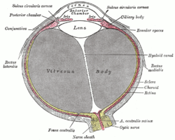
Horizontal section of the eyeball. (Cornea labeled at top, sclera labeled at center right.) Latin tunica fibrosa bulbi, tunica fibrosa oculi Gray's subject #225 1005 The sclera and cornea form the fibrous tunic of the bulb of the eye; the sclera is opaque, and constitutes the posterior five-sixths of the tunic; the cornea is transparent, and forms the anterior sixth.
The term "corneosclera" is also used to describe the sclera and cornea together.[1]
References
- ^ "corneosclera" at Dorland's Medical Dictionary
This article was originally based on an entry from a public domain edition of Gray's Anatomy. As such, some of the information contained within it may be outdated.
Sensory system – visual system – globe of eye (TA A15.2.1–6, TH 3.11.08.0-5, GA 10.1005) Fibrous tunic (outer) Episcleral layer • Schlemm's canal • Trabecular meshworkUvea/vascular tunic (middle) Retina (inner) LayersCellsPhotoreceptor cells (Cone cell, Rod cell) → (Horizontal cell) → Bipolar cell → (Amacrine cell) → Retina ganglion cell (Midget cell, Parasol cell, Bistratified cell, Giant retina ganglion cells, Photosensitive ganglion cell) → Diencephalon: P cell, M cell, K cell
Muller gliaOtherAnterior segment Posterior segment Other M: EYE
anat(g/a/p)/phys/devp/prot
noco/cong/tumr, epon
proc, drug(S1A/1E/1F/1L)
Categories:- Eye anatomy
- Eye stubs
Wikimedia Foundation. 2010.


