- Retinal pigment epithelium
Infobox Anatomy
Name = Retinal pigment epithelium
Latin = p. pigmentosa retinae
GraySubject = 225
GrayPage = 1016
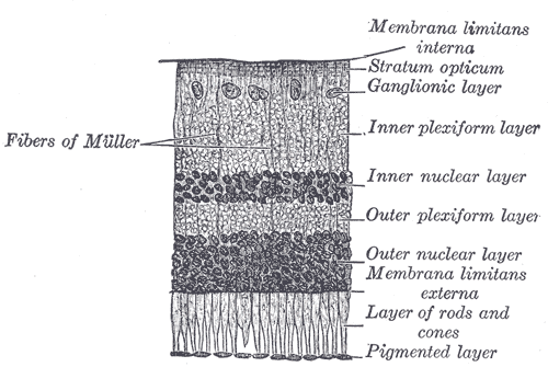
Caption = Section ofretina . (Pigmented layer labeled at bottom right.)
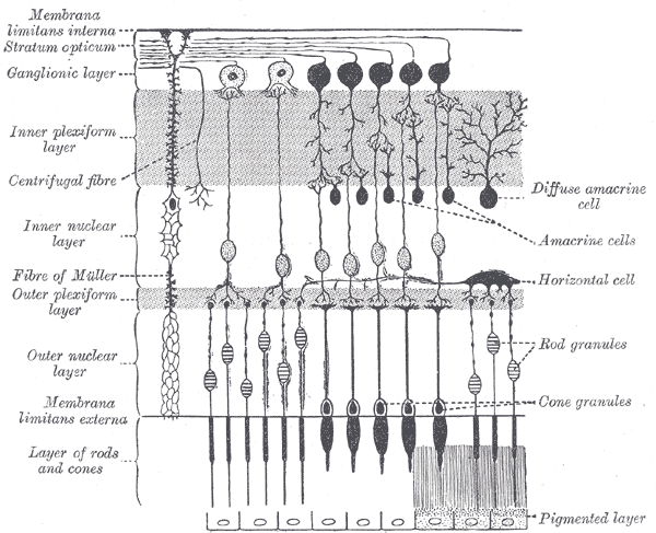
Caption2 = Plan of retinal neurons. (Pigmented layer labeled at bottom right.)
System =
Precursor =
MeshName =
MeshNumber =
DorlandsPre = p_07
DorlandsSuf = 12617535
The retinal pigment epithelium (RPE) is thepigment ed cell layer just outside the neurosensoryretina that nourishes retinal visual cells, and is firmly attached to the underlyingchoroid and overlying retinal visual cells.cite book |author=Cassin, B. and Solomon, S. |title=Dictionary of eye terminology |publisher=Triad Pub. Co |location=Gainesville, Fla |year=2001 |pages= |isbn=0-937404-63-2 |oclc= |doi=] [Boyer MM, Poulsen GL, Nork TM. "Relative contributions of the neurosensory retina and retinal pigment epithelium to macular hypofluorescence." Arch Ophthalmol. 2000 Jan;118(1):27-31. PMID 10636410.]History
The RPE was known in the 18th and 19th centuries as the pigmentum nigrum, referring to the observation that the RPE is dark (black in many animals, brown in humans); and as the tapetum nigrum, referring to the observation that in animals with a
tapetum lucidum , in the region of the tapetum lucidum the RPE is not pigmented. [cite book
author=Coscas, Gabriel and Felice Cardillo Piccolino
title=Retinal Pigment Epithelium and Macular Diseases
publisher=Springer
location=
year=1998
pages= |isbn=0792351444 |oclc= |doi=]Anatomy
The RPE is composed of a single layer of hexagonal cells that are densely packed with pigment granules.
When viewed from the outer surface, these cells are smooth and hexagonal in shape. When seen in section, each cell consists of an outer non-pigmented part containing a large oval nucleus and an inner pigmented portion which extends as a series of straight thread-like processes between the rods, this being especially the case when the eye is exposed to light.
Function
The retinal pigment epithelium is involved in the
phagocytosis of the outer segment of photoreceptor cells and it is also involved in thevitamin A cycle where it isomerizes alltrans retinol to11-cis retinal .The retinal pigment epithelium also serves as the limiting transport factor that maintains the retinal environment by supplying small molecules such as amino acid,
ascorbic acid and D-glucose while remaining a tight barrier to choroidal blood borne substances.Homeostasis of the ionic environment is maintained by a delicate transport exchange system.Pathology
In the eyes of
albinos , the cells of this layer contain no pigment. Dysfunction of the RPE is found inAge-Related Macular Degeneration andRetinitis Pigmentosa .References
ee also
*
Bruch's membrane
*Drusen External links
*MeshName|pigment+epithelium+of+eye
*
*
Wikimedia Foundation. 2010.