- Optic disc
-
Optic disc 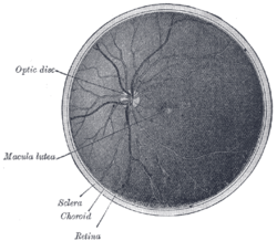
Interior of posterior half of bulb of left eye. The veins are darker in appearance than the arteries. 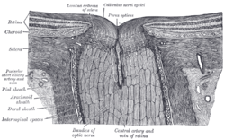
The terminal portion of the optic nerve and its entrance into the eyeball, in horizontal section. Gray's subject #225 1015 MeSH Optic+Disk The optic disc or optic nerve head is the location where ganglion cell axons exit the eye to form the optic nerve. There are no light sensitive rods or cones to respond to a light stimulus at this point. This causes a break in the visual field called "the blind spot" or the "physiological blind spot". The optic disc represents the beginning of the optic nerve (second cranial nerve) and is the point where the axons of retinal ganglion cells come together. The optic disc is also the entry point for the major blood vessels that supply the retina.[1] The optic nerve head in a normal human eye carries from 1 to 1.2 million neurons from the eye towards the brain.
Contents
Anatomy
The optic disc is placed 3 to 4 mm to the nasal side of the fovea. It is a vertical oval, with average dimensions of 1.76mm horizontally by 1.92mm vertically.[2] There is a central depression, of variable size, called the optic cup.
Clinical examination
The eye is unique because of the transparency of its optical media. Almost all eye structures can be examined with appropriate optical equipment and lenses. Using a modern direct ophthalmoscope gives a view of the optic disc using the principle of reversibility of light. A slit lamp biomicroscopic examination along with an appropriate aspheric focusing lens (+66D, +78D or +90D) is required for a detailed stereoscopic view of the optic disc and structures inside the eye.
A biomicroscopic exam can give an indication of the health of the optic nerve. In particular, the eye care physician notes the colour, cupping size (as a cup-to-disc ratio), sharpness of edge, swelling, hemorrhages, notching in the optic disc and any other unusual anomalies. It is useful for finding evidence corroborating the diagnosis of glaucoma and other optic neuropathies, optic neuritis, anterior ischemic optic neuropathy or papilledema (i.e. optic disc swelling produced by raised intracranial pressure), and optic disc drusen.
Women in advanced stage of pregnancy with pre-eclampsia should be screened by an ophthalmoscopic examination of the optic disc for early evidence of rise in intracranial pressure.
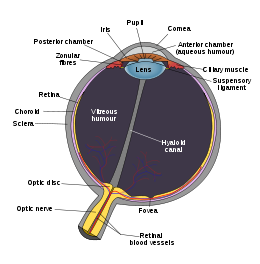 Schematic diagram of the human eye, with the optical disc, or blind spot, at the bottom.
Pale disc
A normal optic disc is orange to pink in colour. A pale disc is an optic disc which varies in colour from a pale pink or orange colour to white. A pale disc is an indication of a disease condition.
Imaging of the optic disc
Traditional colour-film camera images are the gold standard in imaging, requiring an expert ophthalmic photographer, ophthalmic technician, optometrist or an ophthalmologist for taking standardised pictures of the optic disc. Stereoscopic images offer an excellent investigative tool for serial follow-up of suspected changes in the hands of an expert optometrist or ophthalmologist. Automated techniques have also been developed to allow for more efficient and less expensive imaging. Heidelberg Retinal Tomography (HRT-II), GDx-VCC and optical coherence tomography (Stratus-OCT 3) are the currently available computerised techniques for imaging various structures of the eyes, including the optic disc. They quantify the nerve fiber layer of disc and surrounding retina and statistically correlate the findings with a database of previously screened population of normals. They are useful for baseline and serial follow-up to monitor minute changes in optic disc morphology. Imaging will not provide conclusive evidence for clinical diagnosis however, and the evidence needs to be supplanted by serial physiological testing for functional changes. Such tests may include visual field charting, and final clinical interpretation of the complete eye examination by an eye care physician. Ophthalmologists and Optometrists are able to provide this service.
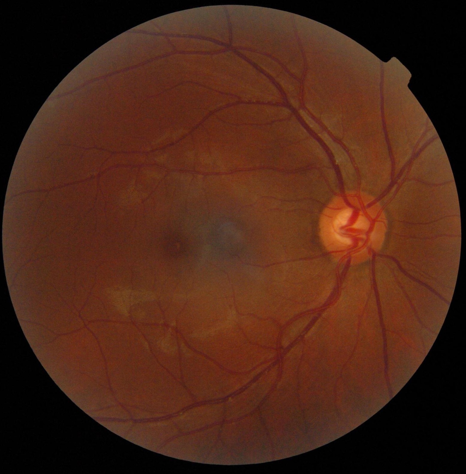
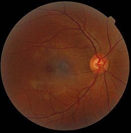 Retinography photograph showing the optic disc as a bright area on the right where blood vessels converge.
References
- ^ blind spot. (2011). In Encyclopædia Britannica. Retrieved from http://www.britannica.com/EBchecked/topic/69390/blind-spot
- ^ Duane's Ophthalmology (2006). Ch. 4 Anatomy of the Visual Sensory System
External links
Sensory system – visual system – globe of eye (TA A15.2.1–6, TH 3.11.08.0-5, GA 10.1005) Fibrous tunic (outer) Episcleral layer • Schlemm's canal • Trabecular meshwork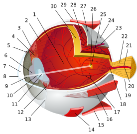

Uvea/vascular tunic (middle) Retina (inner) LayersCellsPhotoreceptor cells (Cone cell, Rod cell) → (Horizontal cell) → Bipolar cell → (Amacrine cell) → Retina ganglion cell (Midget cell, Parasol cell, Bistratified cell, Giant retina ganglion cells, Photosensitive ganglion cell) → Diencephalon: P cell, M cell, K cell
Muller gliaOtherMacula (Foveola, Fovea centralis) • Optic disc (Optic cup)Anterior segment Posterior segment Other M: EYE
anat(g/a/p)/phys/devp/prot
noco/cong/tumr, epon
proc, drug(S1A/1E/1F/1L)
Categories:- Eye anatomy


Wikimedia Foundation. 2010.